
Laboratory Microscopes
实验室显微镜
CLAM · Analytical & Instrumentation · 6 product types · 30 models
By Lab Manufacturers China Technical TeamLast reviewed
What are Laboratory Microscopes?
Laboratory microscopes magnify and resolve fine detail of specimens using transmitted, reflected or fluorescence light. The category covers biological, fluorescence, inverted, metallurgical, polarizing and digital microscopes used in life science, materials, clinical and industrial inspection.
6 product types in Laboratory Microscopes
The 6 product types below are grouped by the exact instrument you need.
- BIM5 models
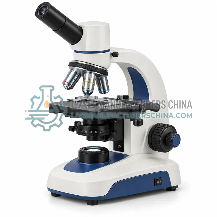
Biological Microscopes
生物显微镜
Biological microscopes examine cells, tissues and microorganisms in transmitted light. They use upright optical paths, interchangeable objectives and contrast methods such as brightfield and phase contrast, and are standard instruments in clinical, microbiology and life-science laboratories.
- FLM5 models
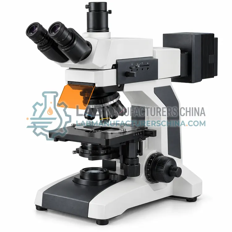
Fluorescence Microscopes
荧光显微镜
Fluorescence microscopes detect light emitted by fluorescently labelled or autofluorescent specimens. They use excitation filters, dichroic mirrors and emission filters with a high-intensity source to reveal specific molecules against a dark background in biological and materials research.
- INM5 models
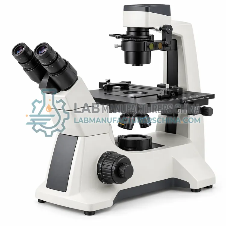
Inverted Microscopes
倒置显微镜
Inverted microscopes place the objective below the stage and the illumination above, so specimens stay in their culture vessel or well. They are used for live-cell culture, tissue work and interaction with samples that cannot be inverted.
- MEM5 models
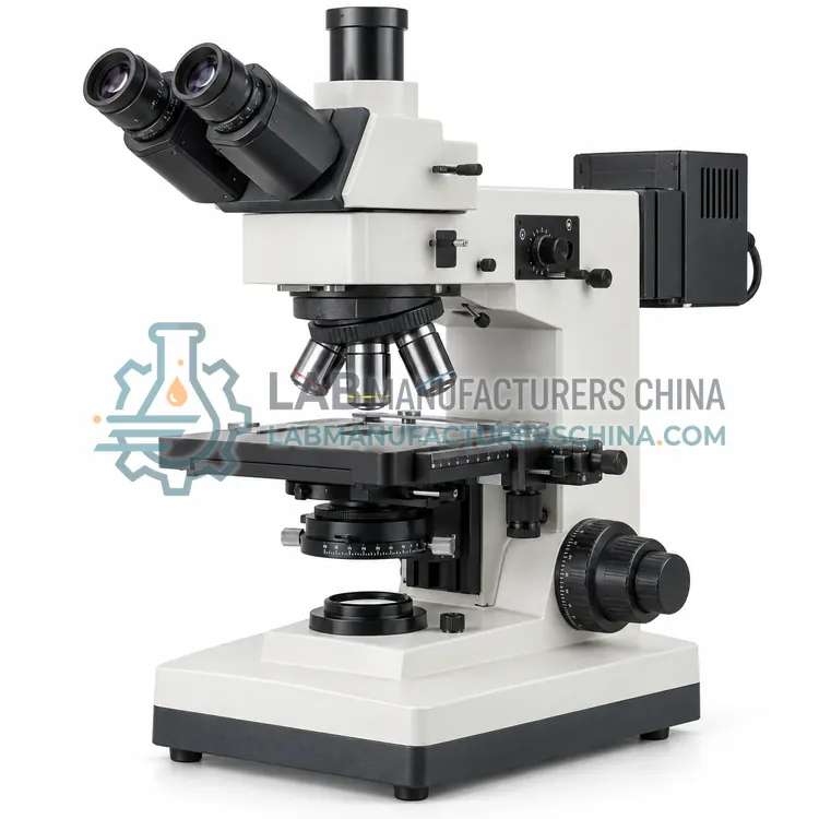
Metallurgical Microscopes
金相显微镜
Metallurgical microscopes examine opaque, reflective specimens in reflected light. They reveal grain structure, phases, inclusions, coatings and defects in metals, alloys, ceramics and electronic components for materials and quality-control laboratories and are central to metallography.
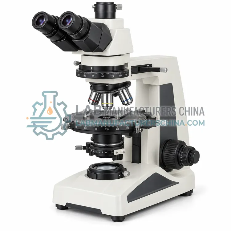
- POM5 models
Polarizing Microscopes
偏光显微镜
Polarizing microscopes use polarized light and a rotating stage to reveal optical anisotropy in crystals, minerals, fibres and polymers. They are used in geology, materials science, forensic and pharmaceutical testing to identify substances and study structure.
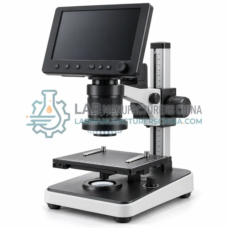
- DIM5 models
Digital Microscopes
数字显微镜
Digital microscopes capture the magnified image with a camera and display it on a screen, replacing or supplementing eyepieces. They support inspection, measurement and documentation with digital images and are used in industry, education and research.
What are Laboratory Microscopes?
Laboratory microscopes are optical instruments that produce a magnified image of a specimen so structures too small for the unaided eye can be examined, measured and documented. The essential elements are an objective lens that forms the magnified image, an eyepiece or camera that further magnifies and presents it, a stage that holds and positions the specimen, and an illumination system matched to the observation method. Different configurations suit different samples: upright biological microscopes view transparent or thinly sectioned specimens in transmitted light, inverted microscopes view living cells from below in culture vessels, metallurgical microscopes view opaque polished surfaces in reflected light, polarizing microscopes reveal optical anisotropy, fluorescence microscopes detect labelled or autofluorescent structures, and digital microscopes capture and analyze images directly. Magnification, numerical aperture, resolution and contrast method determine what can be seen.
How do Laboratory Microscopes work?
The objective lens collects light from the specimen and forms a magnified real image. The eyepiece or tube lens then magnifies this image for the eye or camera. Resolution is set mainly by the numerical aperture of the objective and the wavelength of light, according to the Abbe limit, so a higher numerical aperture resolves finer detail than a larger simple magnification. Contrast methods convert differences in refractive index, thickness or absorption into visible brightness differences: brightfield transmits light directly, darkfield illuminates obliquely, and phase contrast and differential interference contrast enhance transparent specimens. Fluorescence uses excitation filters and dichroic mirrors to illuminate with one wavelength band and detect a longer emitted band. Köhler illumination provides even, controlled lighting.
Where are Laboratory Microscopes used?
Clinical and pathology laboratories use microscopes for blood films, cytology, histology and microbiology. Life science and cell biology laboratories use inverted and fluorescence microscopes for live-cell imaging, immunofluorescence and tissue culture. Materials and metallurgical laboratories examine grain structure, inclusions, coatings and fractures, often with polarized light and reflected illumination. Pharmaceutical laboratories use microscopy for particle characterization, crystal habit and foreign matter identification. Industrial quality departments inspect surfaces, solder joints, fibres and electronic components. Academic and teaching laboratories rely on microscopes for instruction, and digital microscopes support documentation, measurement and remote review of images.
Which standards apply to Laboratory Microscopes?
Microscope optics and accessories are addressed by ISO standards such as ISO 8036 for immersion oil, ISO 19012 for objective designation, ISO 9345 for imaging distances and ISO 8255 for cover glasses. Colorimetric and resolution check methods help verify performance, and stage micrometers and reference specimens are used for calibration of measurement. Fluorescence filter sets are described by their excitation and emission bands. Clinical and biological methods often reference CLSI documents for specimen handling and staining. Electrical safety of laboratory microscopes generally follows IEC 61010-1, and imaging software used in regulated work must support data integrity expectations.
How do I select the right laboratory microscopes equipment?
Start with the specimen and the observation requirement. Transparent or thinly sectioned material suits an upright biological microscope with transmitted brightfield or phase contrast. Living cells in flasks or dishes require an inverted microscope with sufficient working distance. Opaque polished surfaces require a metallurgical microscope with reflected illumination. Specific structures such as crystals, fibres or minerals benefit from polarized light. Fluorescently labelled samples require fluorescence filter sets and a suitable light source. Then choose magnification range, objectives, numerical aperture and contrast method; decide between binocular eyepieces and a camera; and confirm the illumination, stage and software match the workflow. Consider measurement calibration, documentation and regulated data requirements.
Laboratory Microscopes comparison
| Subcategory | Principle | Typical use | Key spec |
|---|---|---|---|
| Biological Microscopes | Transmitted brightfield and contrast methods | Cells, tissues and microbiology | Up to 1000×, NA 1.25 |
| Fluorescence Microscopes | Excitation and emission filter separation | Labelled and autofluorescent structures | Multiple filter cubes, 100 W source |
| Inverted Microscopes | Objective below the stage | Live cells in culture vessels | Long working distance objectives |
| Metallurgical Microscopes | Reflected light from opaque surfaces | Metals, coatings and fractures | Up to 1000×, reflected illumination |
| Polarizing Microscopes | Crossed polarizers for anisotropy | Crystals, minerals and fibres | Rotating stage, strain-free optics |
| Digital Microscopes | Camera and display instead of eyepieces | Documentation, measurement and inspection | 5–20 MP camera, HDMI or USB |
Frequently asked questions about Laboratory Microscopes
Magnification is how much larger the image appears, while resolution is the smallest distance between two points that can still be distinguished as separate. Increasing magnification beyond the limit set by numerical aperture and wavelength produces an empty, blurry image. Resolution therefore matters more than raw magnification.
An inverted microscope is usually chosen for live cells because the objective looks up through the base of the culture flask or dish, leaving space for the vessel and allowing the cells to remain in medium. Incubation chambers and phase contrast or fluorescence accessories support live-cell work.
Numerical aperture describes the range of angles over which an objective can collect light. Higher values collect more light and resolve finer detail, giving brighter and sharper images. It also sets depth of field, so higher numerical aperture gives a shallower in-focus region.
Measurement is calibrated with a stage micrometer of known pitch, typically divided into 10 µm intervals, used to determine the scale for each objective and camera combination. The calibration should be repeated whenever objectives, camera or magnification changers are altered, and recorded for traceability.
Keep optics dust-free with a blower and lens tissue, and never use solvents on cemented lenses or coatings. Check and clean the condenser, filters and light source, lubricate the stage and focus mechanism according to the manual, and store the instrument under a dust cover. Recalibrate after service.
Yes. Many modern microscopes present the image on a monitor through a camera rather than through eyepieces, which reduces eye strain and makes documentation, annotation and measurement easier. Some models retain an eyepiece as well, and the choice depends on the workflow and required image quality.
Related categories
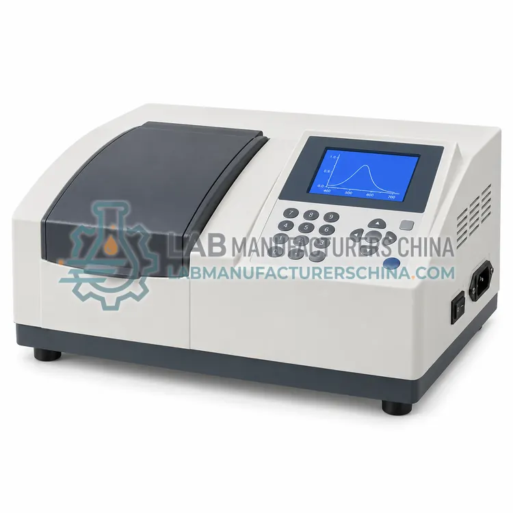 CANI分析仪器Analytical instruments measure the chemical, optical or physical properties of samples to identify substances and quantify their concentration. This category covers spectrophotometers, photometers, refractometers, polarimeters, turbidity meters and general analytical analyzers used in pharmaceutical, environmental, food, chemical and industrial quality-control laboratories.
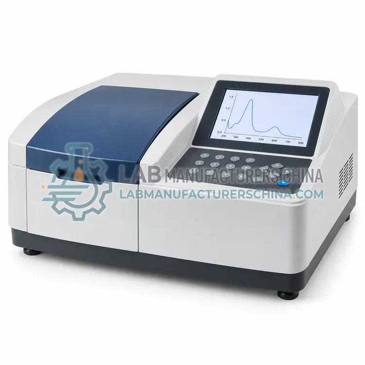 CSPI光谱仪器Spectroscopy instruments measure how matter absorbs, emits or scatters electromagnetic radiation to identify substances and quantify them. This category covers UV-Vis, FTIR, Raman, fluorescence, NIR and atomic spectroscopy systems used in pharmaceutical, chemical, environmental, food and materials laboratories.
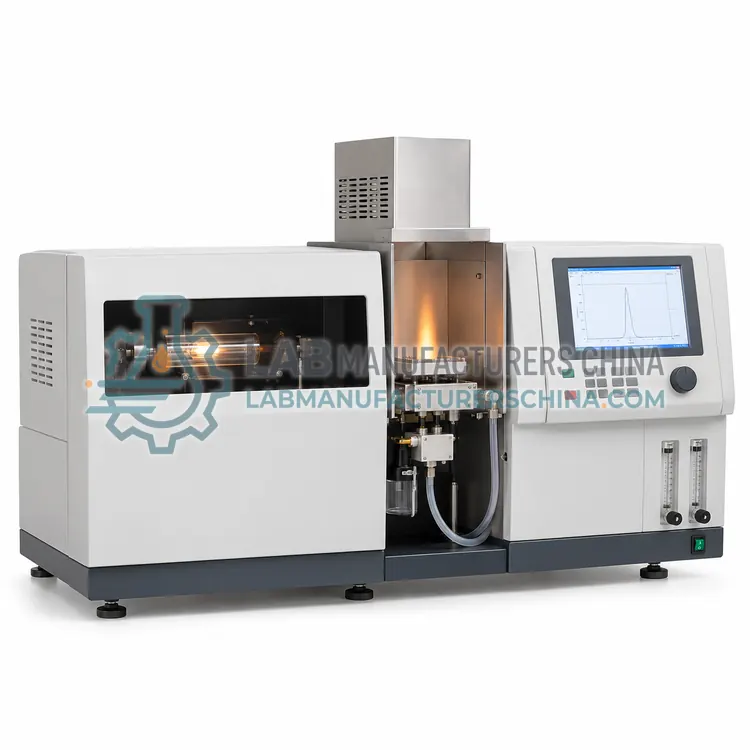 CAAS原子吸收光谱仪Atomic absorption spectrometers determine elemental concentrations by measuring light absorbed by free atoms produced in a flame, graphite furnace or hydride generation cell. They serve environmental, food, clinical, geochemical and pharmaceutical laboratories that need reliable trace-metal results at low cost.
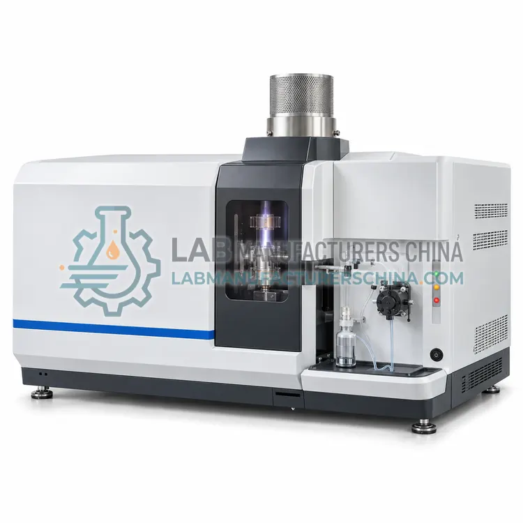 CICSICP 光谱仪ICP spectrometers use an inductively coupled argon plasma to atomize and ionize samples for elemental analysis by optical emission or mass spectrometry. They serve environmental, pharmaceutical, food, metallurgical and geological laboratories that need multi-element results.