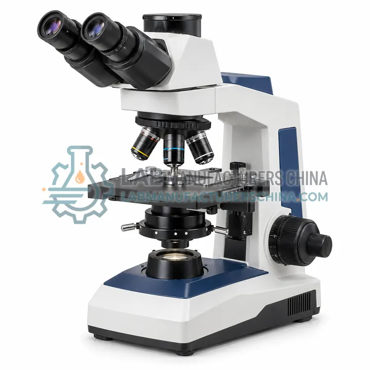
How do Biological Microscopes work?
A biological microscope transmits light from a source through a condenser, the specimen and the objective, then to the eyepiece or camera. The condenser focuses light onto the specimen and, with Köhler alignment, provides even illumination. The objective forms a magnified image whose resolution is limited by its numerical aperture and the light wavelength. A rotating nosepiece brings different objectives into the optical path, and the stage moves the specimen precisely in two axes. Brightfield shows absorption contrast, while phase contrast and darkfield convert refractive index differences into visible contrast for transparent specimens. Focus is adjusted by coarse and fine controls, and total magnification is the product of objective and eyepiece or camera magnification.
Which Biological Microscopes variants are available?
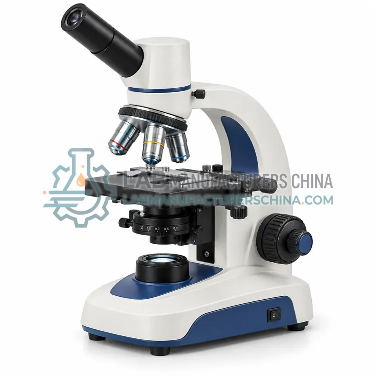
- Monocular Biological Microscope — A monocular biological microscope has a single eyepiece and provides basic transmitted-light observation at low cost.
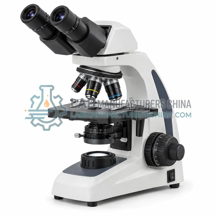
- Binocular Biological Microscope — A binocular biological microscope provides two eyepieces for comfortable routine viewing.
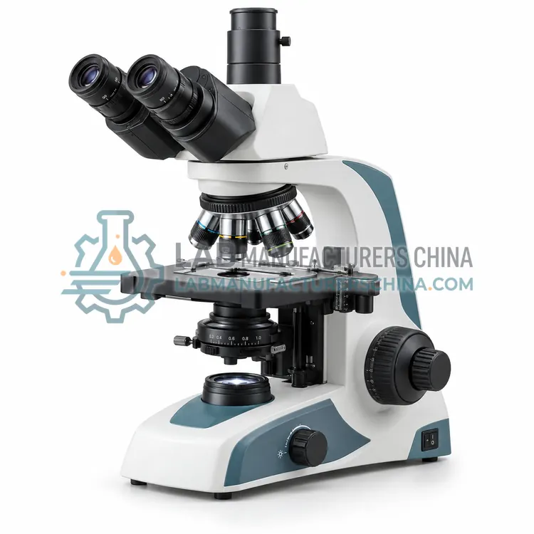
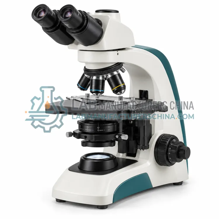
- Trinocular Biological Microscope — A trinocular biological microscope adds a dedicated camera port to a binocular head.
- Phase Contrast Microscope — A phase contrast microscope converts refractive index differences into brightness contrast, making transparent living specimens visible.
- Darkfield Microscope — A darkfield microscope illuminates the specimen obliquely so that only scattered light reaches the objective, showing bright objects on a dark background.
How do I choose the right variant?
Choose a biological microscope for transparent or thinly sectioned specimens in transmitted light. Decide on the observation head first: a monocular head suits basic student use, a binocular head suits routine examination with comfortable viewing, and a trinocular head adds a camera port for documentation. Then select objectives and numerical aperture for the detail required, and add phase contrast or darkfield when unstained living specimens must be examined. Confirm the stage travel, condenser type and illumination source, and whether the eyepieces and objectives match the required field of view. For regulated work, verify measurement calibration and image data handling.
Where are Biological Microscopes used?
Biological Microscopes are specified in clinical cytology and histology, microbiology culture examination, cell and tissue culture research and teaching and student laboratories.
Which standards and certifications apply?
Work with Biological Microscopes is commonly governed by ISO 8036, ISO 19012-1, ISO 9345-1, ISO 8255-1 and IEC 61010-1. Instrument certifications such as CE marking are confirmed per configuration and never assumed.
What are the key considerations before purchase?
Set up Köhler illumination before critical observation so that illumination is even and the numerical aperture of the condenser matches the objective. Keep optics clean with a blower and lens tissue, and avoid solvents on coatings. Use the correct immersion oil with high-numerical-aperture objectives and clean it off after use. Calibrate measurements with a stage micrometer for each objective and camera combination, and record the scale. Store the microscope under a dust cover, and service the focus and stage mechanisms on the schedule recommended by the manufacturer.