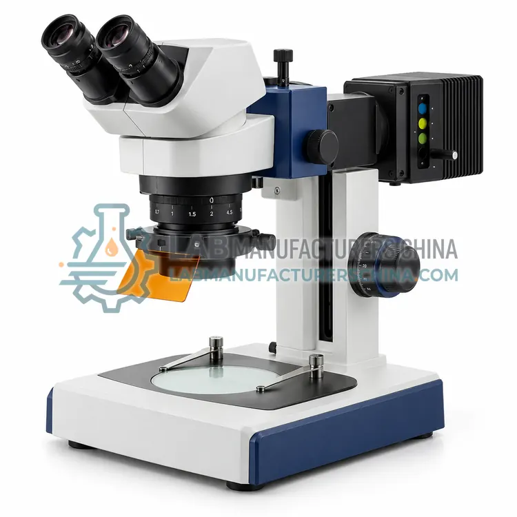
How do Fluorescence Microscopes work?
A fluorescence microscope illuminates the specimen with a selected excitation wavelength using a filter cube containing an excitation filter, a dichroic mirror and an emission filter. Fluorophores in the specimen absorb the excitation light and emit at longer wavelengths. The dichroic mirror reflects the excitation light onto the specimen and transmits the emitted light to the objective and detector, so background illumination is largely excluded. The specimen therefore appears bright against a dark background, giving high contrast and specificity. Multiple filter sets allow different fluorophores to be imaged in turn, and excitation and emission bands must be chosen to avoid overlap between channels. Bright sources, efficient filters and high-numerical-aperture objectives determine sensitivity.
Which Fluorescence Microscopes variants are available?
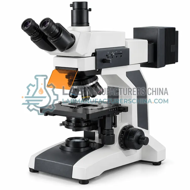
- Upright Fluorescence Microscope — An upright fluorescence microscope illuminates specimens from above through the objective.
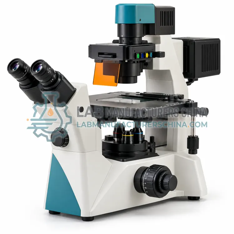
- Inverted Fluorescence Microscope — An inverted fluorescence microscope illuminates and observes live cells from below through the base of the culture vessel.
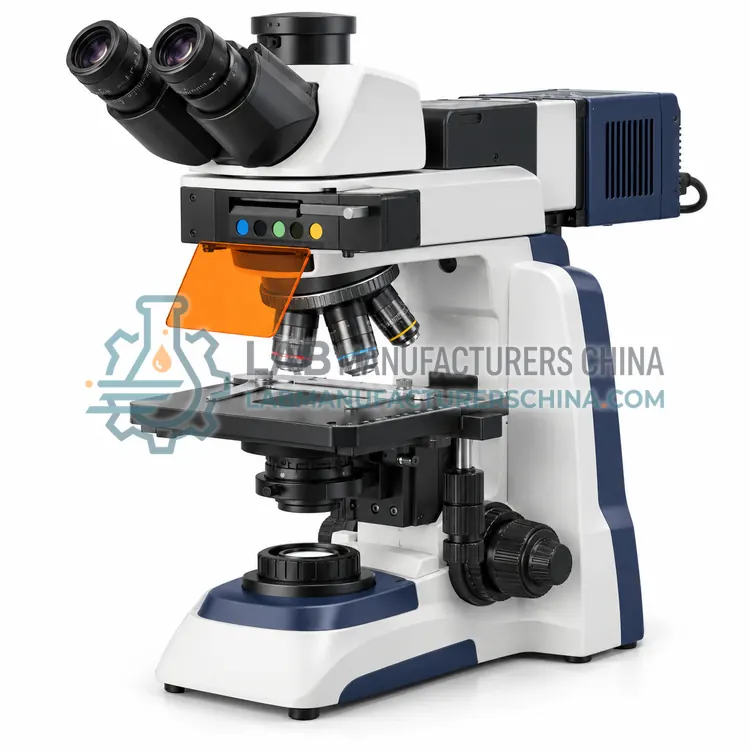
- LED Fluorescence Microscope — An LED fluorescence microscope uses light-emitting diodes instead of arc lamps for excitation.
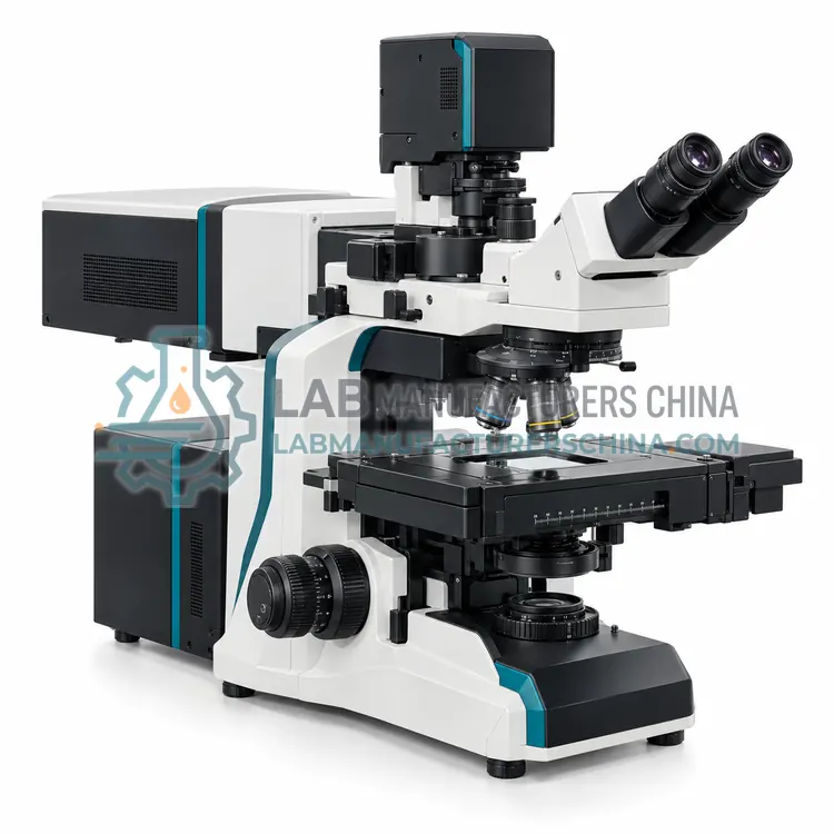
- Confocal Laser Scanning Microscope — A confocal laser scanning microscope uses a focused laser and a pinhole to reject out-of-focus light, giving sharp optical sections.
- Stereo Fluorescence Microscope — A stereo fluorescence microscope combines low-magnification stereoscopic viewing with fluorescence excitation.
How do I choose the right variant?
Choose a fluorescence microscope when specific molecules must be detected through fluorescent labels or natural autofluorescence. Match the filter sets to the fluorophores in use, such as DAPI for nuclei or FITC for green emission, and confirm the excitation source covers all required wavelengths. Decide between an upright configuration for slides and sections and an inverted one for live cells in dishes. For optical sectioning and three-dimensional imaging, a confocal system is required. Consider LED sources for stability and low running cost versus arc lamps for very high intensity. Check objective numerical aperture, transmission and correction, and the camera sensitivity for the emission wavelengths used.
Where are Fluorescence Microscopes used?
Fluorescence Microscopes are specified in immunofluorescence and cell biology, fluorescence in-situ hybridisation, microbiology and viability staining and materials and forensic fluorescence inspection.
Which standards and certifications apply?
Work with Fluorescence Microscopes is commonly governed by ISO 8036, ISO 19012-1, ISO 9345-1, ISO 8255-1 and IEC 61010-1. Instrument certifications such as CE marking are confirmed per configuration and never assumed.
What are the key considerations before purchase?
Protect the specimen from unnecessary light to reduce photobleaching, and use the lowest excitation intensity that gives adequate signal. Select filter sets that match the fluorophores and avoid spectral overlap between channels. Keep filters, dichroic mirrors and objectives clean, since contamination causes background fluorescence and scattering. Mercury and xenon lamps need warm-up, consistent alignment and careful disposal, while light-emitting diodes are stable and long-lived. Verify background and uniformity with a blank slide before imaging. Record excitation and emission settings with the images for reproducibility, and maintain the camera and software for quantitative work.