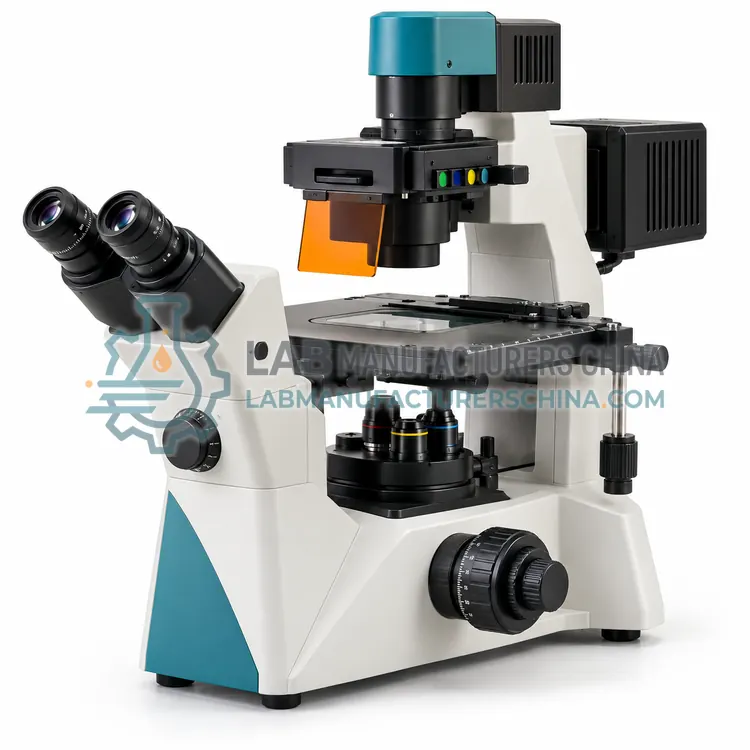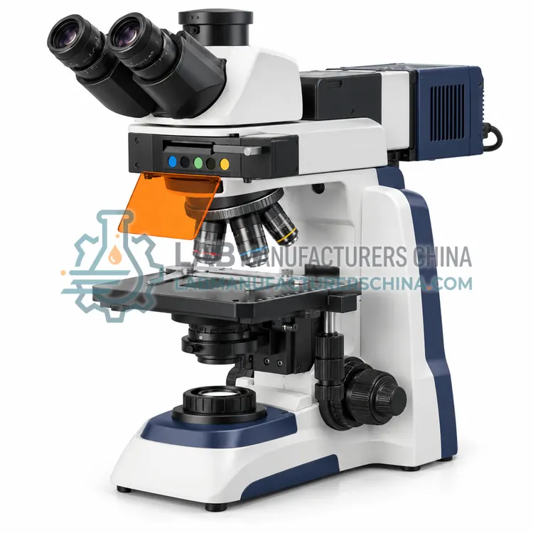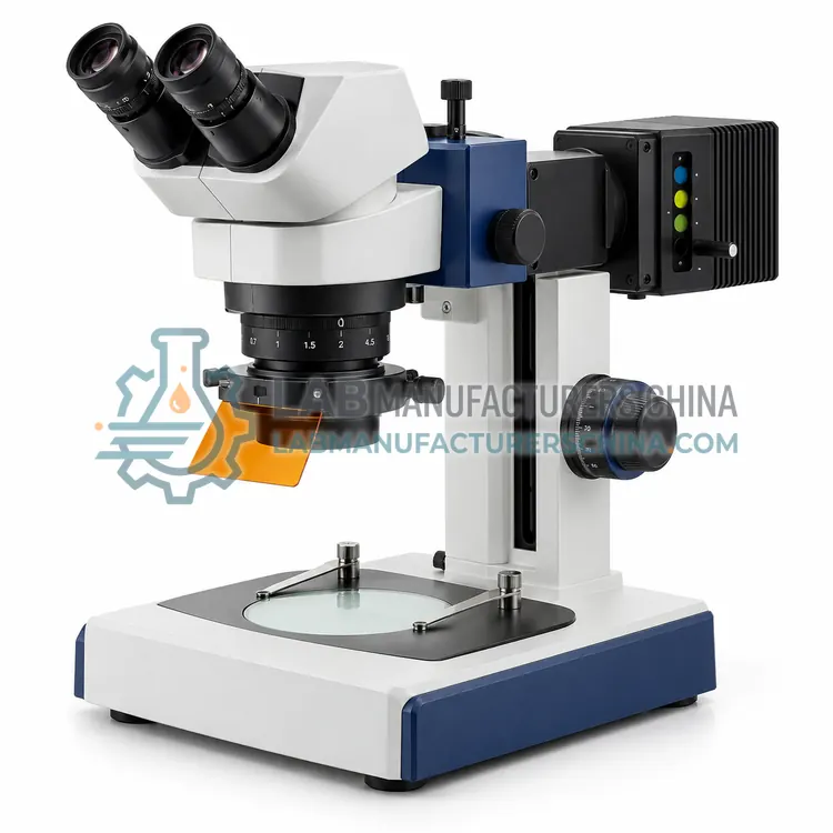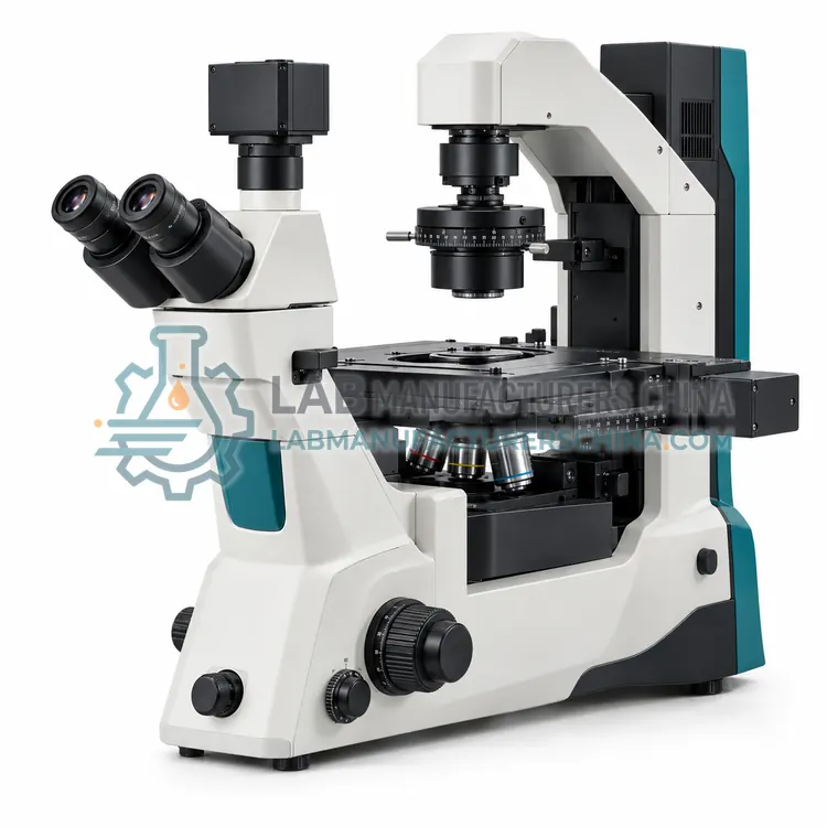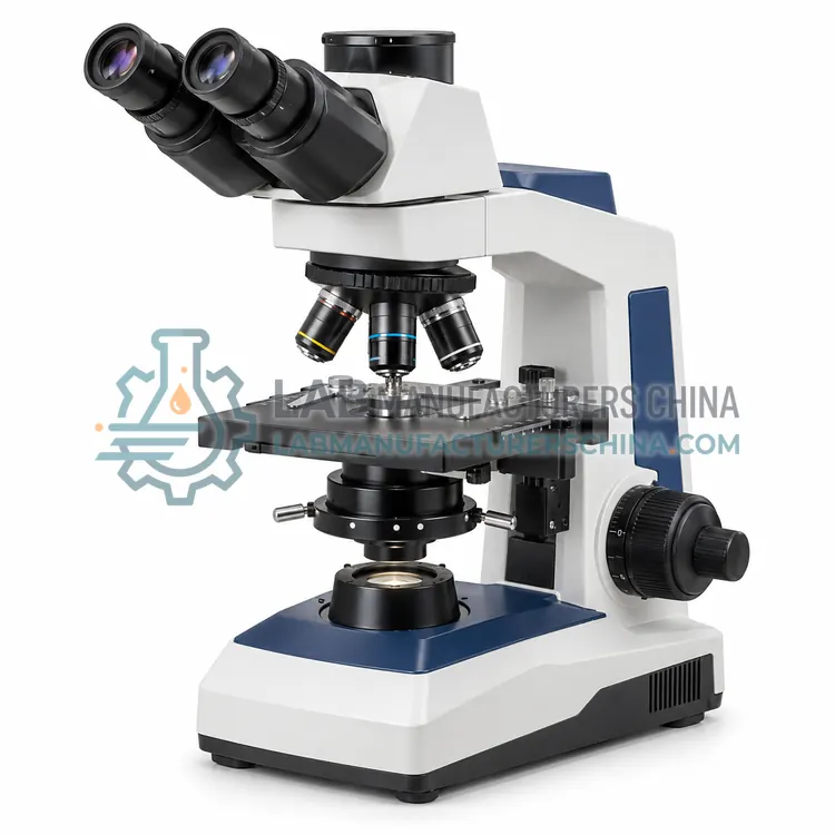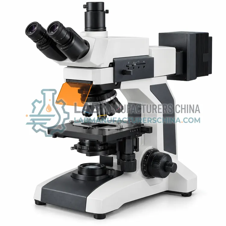
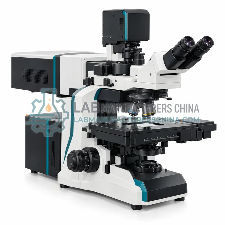
Confocal Laser Scanning Microscope
共聚焦激光扫描显微镜
- SKU FLM-0004
- Fluorescence Microscopes
- CE (to be confirmed)
- ISO 9001 manufacturing (to be confirmed)
Price on request — FOB / CIF quoted per order
Prices depend on configuration, quantity and destination port.
Key specifications
| Laser lines | 405, 488, 561, 640 nm |
|---|---|
| Scan resolution | Up to 4096 × 4096 pixels |
| Detectors | Photomultiplier and GaAsP |
| Pinhole | Adjustable, 1 Airy unit typical |
| Z-step | From 0.1 µm |
| Objectives | 10×, 20×, 40×, 63× oil |
Request a quote Download brochure · to be provided
By Lab Manufacturers China Technical TeamLast reviewed Standards: ISO 8036, ISO 19012-1, ISO 9345-1
What is the Confocal Laser Scanning Microscope?
A confocal laser scanning microscope uses a focused laser and a pinhole to reject out-of-focus light, giving sharp optical sections. It is used for three-dimensional fluorescence imaging of thick specimens and live cells.
What is the Confocal Laser Scanning Microscope?
A confocal laser scanning microscope uses a focused laser and a pinhole to reject out-of-focus light, giving sharp optical sections. It is used for three-dimensional fluorescence imaging of thick specimens and live cells.
Overview
A confocal laser scanning microscope focuses laser light to a diffraction-limited spot on the specimen and scans it point by point across the field. Emitted fluorescence passes back through a pinhole placed at the confocal plane, which blocks light from above and below the focal plane and produces a clean optical section. A photomultiplier or similar detector records the intensity for each point, and the computer builds the image. Scanning the focal plane at successive depths yields a z-stack that can be reconstructed in three dimensions. Multiple laser lines and detectors enable simultaneous or sequential imaging of several fluorophores, and the pinhole size trades resolution and signal against section thickness.
How does it work?
A fluorescence microscope illuminates the specimen with a selected excitation wavelength using a filter cube containing an excitation filter, a dichroic mirror and an emission filter. Fluorophores in the specimen absorb the excitation light and emit at longer wavelengths. The dichroic mirror reflects the excitation light onto the specimen and transmits the emitted light to the objective and detector, so background illumination is largely excluded. The specimen therefore appears bright against a dark background, giving high contrast and specificity. Multiple filter sets allow different fluorophores to be imaged in turn, and excitation and emission bands must be chosen to avoid overlap between channels. Bright sources, efficient filters and high-numerical-aperture objectives determine sensitivity.
What are the key features?
- Laser excitation with point scanning
- Confocal pinhole for optical sectioning
- Z-stack acquisition for three-dimensional reconstruction
- Multiple laser lines and detectors for several fluorophores
- Software for quantitative and time-lapse imaging
What are the technical specifications?
A typical confocal laser scanning microscope configuration offers laser lines of 405, 488, 561, 640 nm, scan resolution of Up to 4096 × 4096 pixels, detectors of Photomultiplier and GaAsP, pinhole of Adjustable, 1 Airy unit typical, Z-step of From 0.1 µm and objectives of 10×, 20×, 40×, 63× oil. Values are indicative of the product class; the exact figures for each configuration are confirmed on the quotation and the calibration documents supplied with the instrument.
Which applications is it used for?
The Confocal Laser Scanning Microscope is used in three-dimensional fluorescence imaging, live-cell and time-lapse confocal imaging, colocalisation studies and optical sectioning of thick tissue. Across the wider Fluorescence Microscopes class, laboratories also deploy it for immunofluorescence and cell biology, fluorescence in-situ hybridisation and microbiology and viability staining.
Which standards and certifications apply?
Relevant standards and methods include ISO 8036, ISO 19012-1, ISO 9345-1, ISO 8255-1 and IEC 61010-1. Certification candidates for this model are CE and ISO 9001 manufacturing; each is to be confirmed for the specific configuration before shipment, and certificates are supplied only where they have been issued.
Protect the specimen from unnecessary light to reduce photobleaching, and use the lowest excitation intensity that gives adequate signal. Select filter sets that match the fluorophores and avoid spectral overlap between channels. Keep filters, dichroic mirrors and objectives clean, since contamination causes background fluorescence and scattering. Mercury and xenon lamps need warm-up, consistent alignment and careful disposal, while light-emitting diodes are stable and long-lived. Verify background and uniformity with a blank slide before imaging. Record excitation and emission settings with the images for reproducibility, and maintain the camera and software for quantitative work.
How do I select the right Confocal Laser Scanning Microscope?
Choose a confocal system when optical sectioning, three-dimensional reconstruction or colocalisation analysis is required. Confirm the laser lines cover the fluorophores, and that detector sensitivity and scan speed suit the sample, especially for live cells. Consider pinhole control, z-stage range and software for acquisition and quantitative analysis. Match objectives to the working distance and correction needed.
How does it compare with other Fluorescence Microscopes variants?
Choose a fluorescence microscope when specific molecules must be detected through fluorescent labels or natural autofluorescence. Match the filter sets to the fluorophores in use, such as DAPI for nuclei or FITC for green emission, and confirm the excitation source covers all required wavelengths. Decide between an upright configuration for slides and sections and an inverted one for live cells in dishes. For optical sectioning and three-dimensional imaging, a confocal system is required. Consider LED sources for stability and low running cost versus arc lamps for very high intensity. Check objective numerical aperture, transmission and correction, and the camera sensitivity for the emission wavelengths used.
Full specifications
| Laser lines | 405, 488, 561, 640 nm |
|---|---|
| Scan resolution | Up to 4096 × 4096 pixels |
| Detectors | Photomultiplier and GaAsP |
| Pinhole | Adjustable, 1 Airy unit typical |
| Z-step | From 0.1 µm |
| Objectives | 10×, 20×, 40×, 63× oil |
| Scan speed | Up to 4 frames per second |
| Stage | Motorised XYZ |
| Image bit depth | 12 or 16 bit |
| Power supply | 100–240 V AC |
Indicative values for this product class. Final specifications are confirmed on the quotation and in the calibration documents supplied with the instrument.
Applications
- Three-dimensional fluorescence imaging
- Live-cell and time-lapse confocal imaging
- Colocalisation studies
- Optical sectioning of thick tissue
Key features
- Laser excitation with point scanning
- Confocal pinhole for optical sectioning
- Z-stack acquisition for three-dimensional reconstruction
- Multiple laser lines and detectors for several fluorophores
- Software for quantitative and time-lapse imaging
Frequently asked questions about Confocal Laser Scanning Microscope
The pinhole blocks light emitted from outside the focal plane, so only the in-focus signal reaches the detector. This produces a thin optical section and improves contrast in thick specimens. A smaller pinhole gives better sectioning but less signal, so size is a trade-off.
Yes, with suitable laser power and scan settings to limit phototoxicity and bleaching. Fast resonant scanners, sensitive detectors and controlled environmental chambers support live-cell confocal imaging, though exposure should be kept as low as possible to preserve cell health.
A z-stack is a series of optical sections acquired at successive focal depths through the specimen. The computer combines them into a three-dimensional reconstruction, which can be viewed as slices, projections or volumes, and used to measure structures in all three dimensions.
A filter cube is a module holding an excitation filter, a dichroic mirror and an emission filter. The excitation filter selects the illumination band, the dichroic mirror reflects it to the specimen and transmits emitted light, and the emission filter blocks residual excitation light before the detector.
Fluorophores can be permanently altered by excitation light, a process called photobleaching. The rate depends on the fluorophore, the excitation intensity and the presence of oxygen. Using lower intensity, shorter exposure, antifade media and efficient filters slows bleaching and preserves the signal.
Standard epifluorescence microscopes image the whole depth of the specimen, so out-of-focus light reduces contrast in thick samples. Optical sectioning requires confocal or light-sheet illumination, which restricts detection to a thin focal plane and enables three-dimensional reconstruction.
Related models