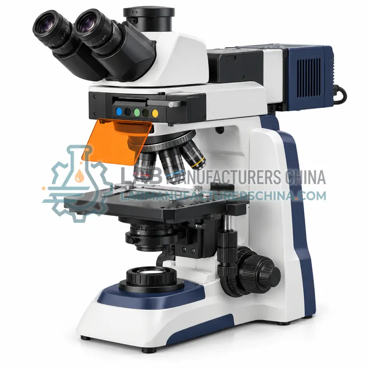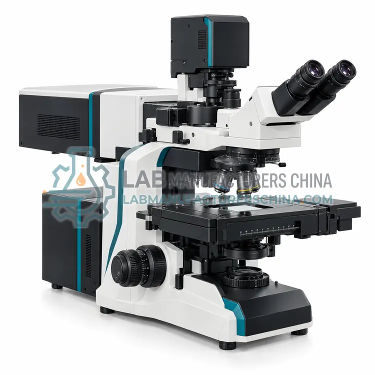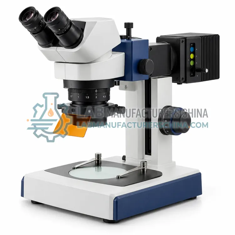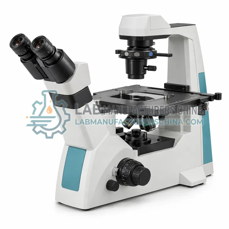
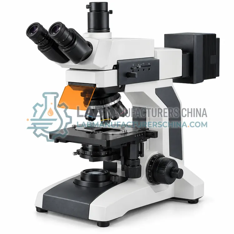
Upright Fluorescence Microscope
正置荧光显微镜
- SKU FLM-0001
- Fluorescence Microscopes
- CE (to be confirmed)
- ISO 9001 manufacturing (to be confirmed)
Price on request — FOB / CIF quoted per order
Prices depend on configuration, quantity and destination port.
Key specifications
| Optical system | Infinite corrected, fluorescence |
|---|---|
| Filter cubes | DAPI, FITC, TRITC, 4-position turret |
| Light source | 100 W mercury lamp or LED |
| Objectives | 10×, 20×, 40×, 100× semi-apochromat |
| Eyepiece | 10× widefield |
| Stage | Mechanical, 140 × 140 mm |
Request a quote Download brochure · to be provided
By Lab Manufacturers China Technical TeamLast reviewed Standards: ISO 8036, ISO 19012-1, ISO 9345-1
What is the Upright Fluorescence Microscope?
An upright fluorescence microscope illuminates specimens from above through the objective. It is the standard configuration for fluorescently labelled tissue sections, smears and fixed cells that are prepared and mounted on glass slides.
What is the Upright Fluorescence Microscope?
An upright fluorescence microscope illuminates specimens from above through the objective. It is the standard configuration for fluorescently labelled tissue sections, smears and fixed cells that are prepared and mounted on glass slides.
Overview
An upright fluorescence microscope uses episcopic illumination, in which the excitation light passes through the objective to the specimen and the emitted light returns through the same objective. This design keeps the objective close to the specimen for high numerical aperture and resolution. Slides sit on a mechanical stage above the condenser position, and transmitted illumination is available for brightfield comparison. Multiple filter cubes in a turret allow rapid switching between fluorophores, and the trinocular head accepts a camera. Arc lamps or light-emitting diodes provide excitation. The configuration is ideal for mounted specimens, sections and smears, where the sample is thin and prepared on a flat surface.
How does it work?
A fluorescence microscope illuminates the specimen with a selected excitation wavelength using a filter cube containing an excitation filter, a dichroic mirror and an emission filter. Fluorophores in the specimen absorb the excitation light and emit at longer wavelengths. The dichroic mirror reflects the excitation light onto the specimen and transmits the emitted light to the objective and detector, so background illumination is largely excluded. The specimen therefore appears bright against a dark background, giving high contrast and specificity. Multiple filter sets allow different fluorophores to be imaged in turn, and excitation and emission bands must be chosen to avoid overlap between channels. Bright sources, efficient filters and high-numerical-aperture objectives determine sensitivity.
What are the key features?
- Episcopic illumination through the objective
- Turret for multiple fluorescence filter cubes
- Mechanical stage for prepared slides
- Trinocular head for camera attachment
- Transmitted light option for brightfield comparison
What are the technical specifications?
A typical upright fluorescence microscope configuration offers optical system of Infinite corrected, fluorescence, filter cubes of DAPI, FITC, TRITC, 4-position turret, light source of 100 W mercury lamp or LED, objectives of 10×, 20×, 40×, 100× semi-apochromat, eyepiece of 10× widefield and stage of Mechanical, 140 × 140 mm. Values are indicative of the product class; the exact figures for each configuration are confirmed on the quotation and the calibration documents supplied with the instrument.
Which applications is it used for?
The Upright Fluorescence Microscope is used in immunofluorescence on tissue sections, fluorescence in-situ hybridisation, smear and cytology fluorescence and microbiology fluorescence staining. Across the wider Fluorescence Microscopes class, laboratories also deploy it for immunofluorescence and cell biology, fluorescence in-situ hybridisation and microbiology and viability staining.
Which standards and certifications apply?
Relevant standards and methods include ISO 8036, ISO 19012-1, ISO 9345-1, ISO 8255-1 and IEC 61010-1. Certification candidates for this model are CE and ISO 9001 manufacturing; each is to be confirmed for the specific configuration before shipment, and certificates are supplied only where they have been issued.
Protect the specimen from unnecessary light to reduce photobleaching, and use the lowest excitation intensity that gives adequate signal. Select filter sets that match the fluorophores and avoid spectral overlap between channels. Keep filters, dichroic mirrors and objectives clean, since contamination causes background fluorescence and scattering. Mercury and xenon lamps need warm-up, consistent alignment and careful disposal, while light-emitting diodes are stable and long-lived. Verify background and uniformity with a blank slide before imaging. Record excitation and emission settings with the images for reproducibility, and maintain the camera and software for quantitative work.
How do I select the right Upright Fluorescence Microscope?
Select an upright fluorescence microscope for thin, prepared specimens on slides. Match the filter cubes to the fluorophores and confirm the source covers all required wavelengths. Choose objectives with high numerical aperture and good transmission at the emission wavelengths, and a camera with adequate sensitivity for the signal level. Add transmitted illumination if brightfield comparison is needed.
How does it compare with other Fluorescence Microscopes variants?
Choose a fluorescence microscope when specific molecules must be detected through fluorescent labels or natural autofluorescence. Match the filter sets to the fluorophores in use, such as DAPI for nuclei or FITC for green emission, and confirm the excitation source covers all required wavelengths. Decide between an upright configuration for slides and sections and an inverted one for live cells in dishes. For optical sectioning and three-dimensional imaging, a confocal system is required. Consider LED sources for stability and low running cost versus arc lamps for very high intensity. Check objective numerical aperture, transmission and correction, and the camera sensitivity for the emission wavelengths used.
Full specifications
| Optical system | Infinite corrected, fluorescence |
|---|---|
| Filter cubes | DAPI, FITC, TRITC, 4-position turret |
| Light source | 100 W mercury lamp or LED |
| Objectives | 10×, 20×, 40×, 100× semi-apochromat |
| Eyepiece | 10× widefield |
| Stage | Mechanical, 140 × 140 mm |
| Illumination modes | Episcopic fluorescence and transmitted |
| Camera port | C-mount trinocular |
| Focus adjustment | Coarse and fine |
| Power supply | 100–240 V AC |
Indicative values for this product class. Final specifications are confirmed on the quotation and in the calibration documents supplied with the instrument.
Applications
- Immunofluorescence on tissue sections
- Fluorescence in-situ hybridisation
- Smear and cytology fluorescence
- Microbiology fluorescence staining
Key features
- Episcopic illumination through the objective
- Turret for multiple fluorescence filter cubes
- Mechanical stage for prepared slides
- Trinocular head for camera attachment
- Transmitted light option for brightfield comparison
Frequently asked questions about Upright Fluorescence Microscope
In episcopic illumination the objective acts as both condenser and collector, so excitation and emission share the same optical path. This allows high-numerical-aperture objectives to be used and keeps the optical axis simple, which is efficient for capturing the weak emitted light from a small area.
It can image live cells grown on glass coverslips or in shallow chambers, but bathing medium and the vessel geometry make an inverted microscope more convenient for cells in flasks, dishes or multi-well plates. The upright design is best for mounted, flat specimens.
Most work needs two to four cubes, corresponding to the fluorophores in use, such as blue, green and red channels. A turret holding several cubes allows quick changes without removing the objective or disturbing the specimen, which helps preserve focus and reduce bleaching.
A filter cube is a module holding an excitation filter, a dichroic mirror and an emission filter. The excitation filter selects the illumination band, the dichroic mirror reflects it to the specimen and transmits emitted light, and the emission filter blocks residual excitation light before the detector.
Fluorophores can be permanently altered by excitation light, a process called photobleaching. The rate depends on the fluorophore, the excitation intensity and the presence of oxygen. Using lower intensity, shorter exposure, antifade media and efficient filters slows bleaching and preserves the signal.
Standard epifluorescence microscopes image the whole depth of the specimen, so out-of-focus light reduces contrast in thick samples. Optical sectioning requires confocal or light-sheet illumination, which restricts detection to a thin focal plane and enables three-dimensional reconstruction.
Related models