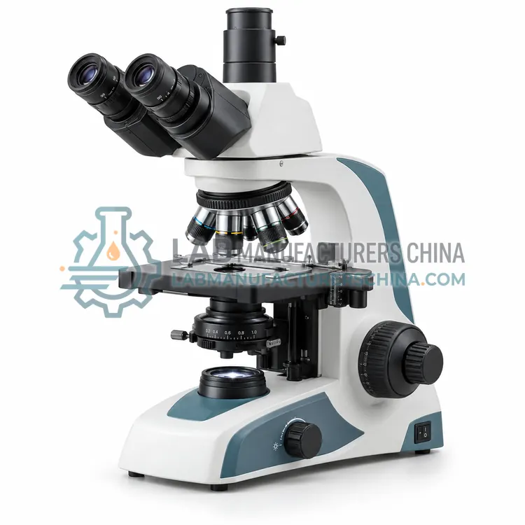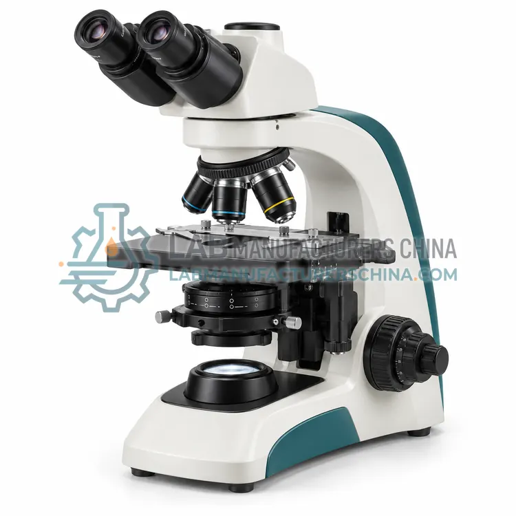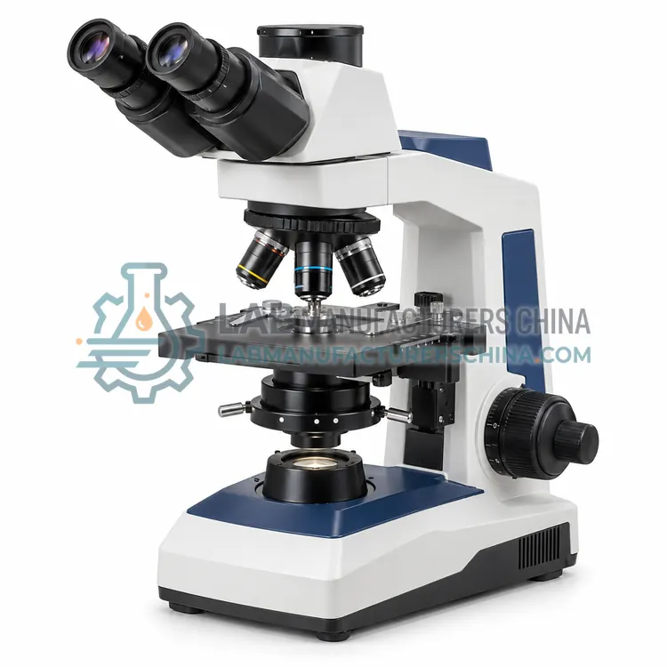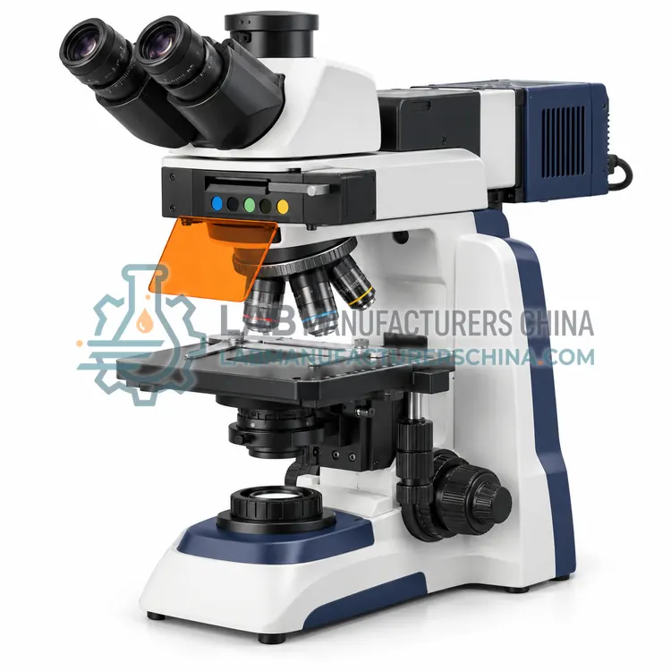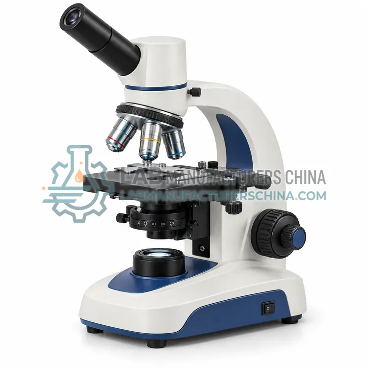
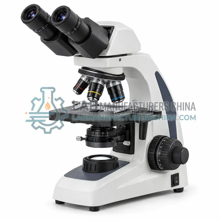
Binocular Biological Microscope
双目生物显微镜
- SKU BIM-0002
- Biological Microscopes
- CE (to be confirmed)
- ISO 9001 manufacturing (to be confirmed)
Price on request — FOB / CIF quoted per order
Prices depend on configuration, quantity and destination port.
Key specifications
| Optical system | Infinite corrected |
|---|---|
| Magnification range | 40×–1000× |
| Objectives | 4×, 10×, 40×, 100× plan |
| Eyepiece | 10× widefield binocular |
| Interpupillary range | 48–75 mm |
| Stage | Mechanical, 140 × 140 mm |
Request a quote Download brochure · to be provided
By Lab Manufacturers China Technical TeamLast reviewed Standards: ISO 8036, ISO 19012-1, ISO 9345-1
What is the Binocular Biological Microscope?
A binocular biological microscope provides two eyepieces for comfortable routine viewing. It is the standard laboratory instrument for cytology, microbiology and histology, balancing optical performance, ergonomics and cost for busy laboratories that examine many specimens.
What is the Binocular Biological Microscope?
A binocular biological microscope provides two eyepieces for comfortable routine viewing. It is the standard laboratory instrument for cytology, microbiology and histology, balancing optical performance, ergonomics and cost for busy laboratories that examine many specimens.
Overview
A binocular biological microscope presents the magnified image to both eyes through a prism assembly that splits the light path. Interpupillary distance and diopter adjustments match the user's eyes, reducing fatigue during long sessions. Plan objectives correct field curvature for sharp images across the whole field, and a five-position nosepiece speeds objective changes. Köhler illumination and an Abbe or achromatic condenser provide even light and controllable contrast. The mechanical stage allows smooth two-axis positioning, and the frame is rigid enough to hold alignment. This configuration is the workhorse of clinical and research laboratories. A trinocular port is offered on similar models when imaging is needed.
How does it work?
A biological microscope transmits light from a source through a condenser, the specimen and the objective, then to the eyepiece or camera. The condenser focuses light onto the specimen and, with Köhler alignment, provides even illumination. The objective forms a magnified image whose resolution is limited by its numerical aperture and the light wavelength. A rotating nosepiece brings different objectives into the optical path, and the stage moves the specimen precisely in two axes. Brightfield shows absorption contrast, while phase contrast and darkfield convert refractive index differences into visible contrast for transparent specimens. Focus is adjusted by coarse and fine controls, and total magnification is the product of objective and eyepiece or camera magnification.
What are the key features?
- Two eyepieces with interpupillary and diopter adjustment
- Plan objectives for flat fields of view
- Five-position nosepiece for quick objective changes
- Köhler illumination with achromatic condenser
- Rigid stand for stable alignment
What are the technical specifications?
A typical binocular biological microscope configuration offers optical system of Infinite corrected, magnification range of 40×–1000×, objectives of 4×, 10×, 40×, 100× plan, eyepiece of 10× widefield binocular, interpupillary range of 48–75 mm and stage of Mechanical, 140 × 140 mm. Values are indicative of the product class; the exact figures for each configuration are confirmed on the quotation and the calibration documents supplied with the instrument.
Which applications is it used for?
The Binocular Biological Microscope is used in clinical cytology and haematology, microbiology and bacteriology, histology and pathology and pharmaceutical and food microscopy. Across the wider Biological Microscopes class, laboratories also deploy it for clinical cytology and histology, microbiology culture examination and cell and tissue culture research.
Which standards and certifications apply?
Relevant standards and methods include ISO 8036, ISO 19012-1, ISO 9345-1, ISO 8255-1 and IEC 61010-1. Certification candidates for this model are CE and ISO 9001 manufacturing; each is to be confirmed for the specific configuration before shipment, and certificates are supplied only where they have been issued.
Set up Köhler illumination before critical observation so that illumination is even and the numerical aperture of the condenser matches the objective. Keep optics clean with a blower and lens tissue, and avoid solvents on coatings. Use the correct immersion oil with high-numerical-aperture objectives and clean it off after use. Calibrate measurements with a stage micrometer for each objective and camera combination, and record the scale. Store the microscope under a dust cover, and service the focus and stage mechanisms on the schedule recommended by the manufacturer.
How do I select the right Binocular Biological Microscope?
Choose a binocular microscope for routine examination where comfortable, sustained viewing is required. Confirm that the objectives include plan versions if sharpness across the field matters, and that the condenser suits brightfield and any add-on contrast method. Check interpupillary and diopter adjustment for the users, and the durability of the stage for daily use.
How does it compare with other Biological Microscopes variants?
Choose a biological microscope for transparent or thinly sectioned specimens in transmitted light. Decide on the observation head first: a monocular head suits basic student use, a binocular head suits routine examination with comfortable viewing, and a trinocular head adds a camera port for documentation. Then select objectives and numerical aperture for the detail required, and add phase contrast or darkfield when unstained living specimens must be examined. Confirm the stage travel, condenser type and illumination source, and whether the eyepieces and objectives match the required field of view. For regulated work, verify measurement calibration and image data handling.
Full specifications
| Optical system | Infinite corrected |
|---|---|
| Magnification range | 40×–1000× |
| Objectives | 4×, 10×, 40×, 100× plan |
| Eyepiece | 10× widefield binocular |
| Interpupillary range | 48–75 mm |
| Stage | Mechanical, 140 × 140 mm |
| Illumination | 3 W LED, brightness adjustable |
| Condenser | Abbe NA 1.25 with iris |
| Nosepiece | 5-position |
| Power supply | 100–240 V AC |
Indicative values for this product class. Final specifications are confirmed on the quotation and in the calibration documents supplied with the instrument.
Applications
- Clinical cytology and haematology
- Microbiology and bacteriology
- Histology and pathology
- Pharmaceutical and food microscopy
Key features
- Two eyepieces with interpupillary and diopter adjustment
- Plan objectives for flat fields of view
- Five-position nosepiece for quick objective changes
- Köhler illumination with achromatic condenser
- Rigid stand for stable alignment
Frequently asked questions about Binocular Biological Microscope
Two-eyed viewing allows the brain to fuse the two images, which reduces fatigue and improves depth perception and comfort during long sessions. For clinical and research work where many specimens are examined daily, a binocular head is a practical necessity rather than a luxury.
Plan objectives are corrected for field curvature, so the image stays sharp from the centre to the edge of the field. Standard achromatic objectives may blur near the edges. Plan optics are preferred for photography and for examining structures across the entire field.
The eyepieces slide or rotate to match the distance between the user's pupils, usually between about 48 and 75 mm, until the two images merge into one circular field. A diopter ring on one eyepiece then corrects any difference in eye focus between the two eyes.
A monocular head has a single eyepiece and is economical for teaching. A binocular head has two eyepieces for comfortable routine viewing. A trinocular head adds a third port for a camera, so it is preferred when images must be captured or shared.
Phase contrast is used to view transparent, unstained specimens such as living cells and microorganisms, which show little contrast in brightfield. It converts differences in refractive index into brightness differences, making outlines and internal structures visible without staining or fixation.
Immersion oil has a refractive index close to that of glass and is placed between the specimen coverslip and a high-numerical-aperture objective. It prevents light loss at the air gap, allowing the objective to reach its rated numerical aperture and resolution. It must be cleaned off after use.
Related models