
Histology Equipment
组织学设备
CHIE · Clinical & Diagnostic · 5 product types · 25 models
By Lab Manufacturers China Technical TeamLast reviewed
What are Histology Equipment?
Histology equipment covers the instruments used to embed, section, float, dry and stain tissue samples for microscopic examination. It serves hospital pathology laboratories, research histology facilities and contract research organizations that turn biopsy and necropsy specimens into stained slides for diagnosis and study.
5 product types in Histology Equipment
The 5 product types below are grouped by the exact instrument you need.
- RMI5 models
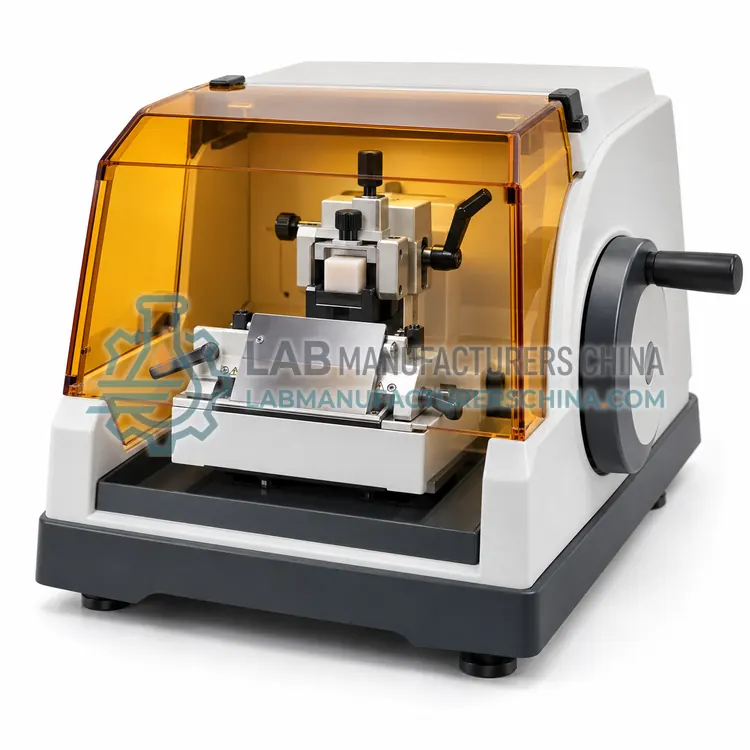
Rotary Microtomes
轮转式切片机
Rotary microtomes are the standard sectioning instruments in histology, using a handwheel or motor-driven block advance to cut consistent paraffin sections from 1 to 60 µm. Pathology and research laboratories choose them for routine biopsy work, serial sectioning and special stains.
- CRY5 models
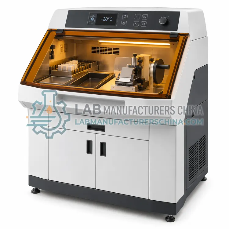
Cryostats
恒冷箱切片机
Cryostats are refrigerated microtomes that cut frozen tissue sections at about –20 °C, used for intraoperative diagnosis and for fresh tissue that must not be chemically processed. They serve surgical pathology, immunohistochemistry and research laboratories that need rapid or native-state sections.
- TFB5 models
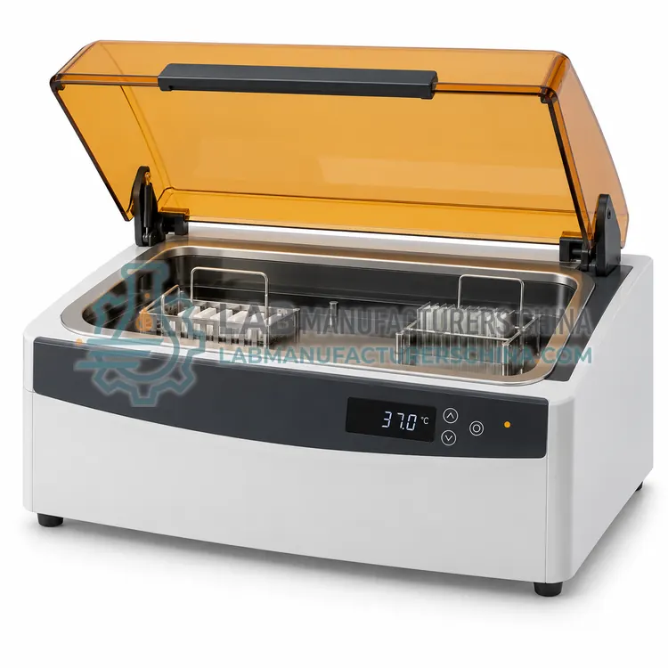
Tissue Float Baths
展片水浴
Tissue float baths are shallow heated water baths used to stretch paraffin section ribbons flat before they are picked up on glass slides. Histology and pathology laboratories rely on them to remove wrinkles and folds so sections stain evenly and can be read under the microscope.
- SLD5 models
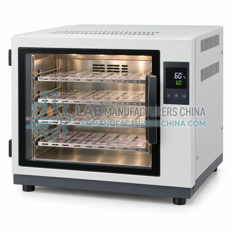
Slide Dryers
烤片器
Slide dryers are heated plates or ovens that warm mounted sections so residual paraffin melts and the tissue adheres firmly to the glass. Histology and pathology laboratories use them before staining to prevent sections detaching during processing.
- HIS5 models
Histology Stainers
组织学染色机
Histology stainers apply hematoxylin and eosin or special stains to tissue sections by moving slide racks through timed reagent stations. Pathology and research laboratories use them to obtain consistent colour and reproducible staining across large daily workloads.
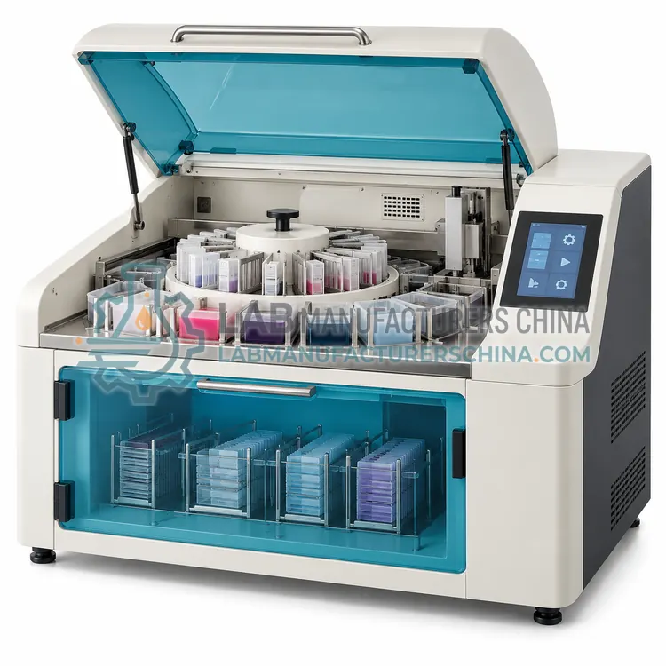
What are Histology Equipment?
Histology equipment is the workflow chain that converts a fixed tissue specimen into a stained, coverslipped slide a pathologist can read under a microscope. Rotary microtomes and cryostats cut the sections; tissue float baths flatten them onto glass; slide dryers bond them to the glass; and stainers apply hematoxylin and eosin or special stains. Around these core steps sit tissue processors and embedding stations for paraffin work, plus blades, cassettes, molds and slides. Each instrument must deliver consistent section thickness, temperature or staining time, because diagnostic decisions rest on reproducible morphology, and laboratories usually document and validate performance under quality-system requirements.
How do Histology Equipment work?
Mechanically, a rotary microtome advances the paraffin block by a set increment, usually 1 to 60 µm, while a handwheel drives the block past a fixed blade to form a ribbon of sections. A cryostat does the same inside a chamber held near –20 °C so fresh tissue can be cut for rapid diagnosis. Float baths heat demineralised or purified water to about 40 to 45 °C so the ribbon stretches flat by surface tension before it is picked up on a slide. Slide dryers hold 55 to 65 °C to melt residual paraffin and adhere the section. Stainers move slide racks through timed reagent stations, then a coverslipper applies resin or film. Temperature, timing and fluid purity are the parameters that determine the result.
Where are Histology Equipment used?
Histology equipment is used wherever tissue must be examined structurally. Hospital pathology departments rely on processors, microtomes and stainers for surgical biopsy and resection reporting, while cryostats support intraoperative frozen sections during surgery. Research institutes and pharmaceutical companies use the same instruments for toxicology, neuroscience, oncology and developmental studies, often with special stains, immunohistochemistry or in-situ hybridisation. Veterinary and agricultural laboratories apply it to animal and plant specimens. Forensic and materials laboratories also section embedded samples. The common requirement is reproducible morphology and traceable records from accession to stained slide.
Which standards apply to Histology Equipment?
Laboratory practice is usually governed by ISO 15189 for medical laboratories and ISO 9001 for quality management, with national pathology guidelines adding detail. Instrument safety follows IEC 61010-1 and, for laboratory equipment, IEC 61010-2-101 and IEC 61326-1 for EMC. Manufacturer quality systems follow ISO 13485 where the device is regulated as in-vitro diagnostic equipment, and CE marking or FDA listing may apply in some markets. Tissue processing and staining reagents are covered by pharmacopoeial and safety regulations. Procurement should confirm that validation, calibration and maintenance documents accompany each instrument, and any certification claims remain to be confirmed per model.
How do I select the right histology equipment?
Select histology equipment by working backwards from the specimen and the report you must issue. For routine paraffin work, choose a processor and an automatic or semi-automatic rotary microtome rated for your block throughput, then match float bath and slide dryer sizes to the same batch volume. Add a cryostat if frozen sections are needed, and pick staining capacity in slides per run with the reagent menu you actually use. Check section thickness range and increment, temperature uniformity, warm-up time, chemical resistance of wetted parts, and compatibility of cassettes and molds. Confirm consumable supply, service response, training and validation support, since these determine long-term cost and compliance more than purchase price.
Histology Equipment comparison
| Subcategory | Principle | Typical use | Key spec |
|---|---|---|---|
| Rotary Microtomes | Handwheel drives block past a fixed blade with micrometer feed | Routine paraffin sections for diagnosis | Section thickness 1–60 µm; increment 1 µm |
| Cryostats | Microtome inside a –20 °C refrigerated chamber | Intraoperative frozen sections and fresh tissue | Chamber –20 to –30 °C; thickness 1–100 µm |
| Tissue Float Baths | Heated water bath stretches section ribbon by surface tension | Flattening paraffin ribbons before mounting | Bath 40–50 °C; control ±1 °C |
| Slide Dryers | Conductive hotplate or oven adheres sections to glass | Baking slides before staining | Temperature 55–65 °C; capacity in slides |
| Histology Stainers | Timed transfer of slide racks through reagent stations | H&E and special stains for tissue slides | Throughput 20–500 slides per run |
Frequently asked questions about Histology Equipment
A histology laboratory typically uses tissue processors, embedding stations, rotary microtomes, float baths, slide dryers and stainers, plus a cryostat for frozen sections and a coverslipper. The exact set depends on whether the laboratory does routine paraffin work, frozen sections or research staining.
A microtome cuts paraffin-embedded or fixed tissue at room temperature, while a cryostat contains a microtome inside a refrigerated chamber at about –20 °C and cuts fresh frozen tissue. Cryostats are used when a rapid answer is needed, such as intraoperative diagnosis.
The flotation bath is warmed to about 40 to 45 °C, which softens the paraffin and lets surface tension stretch the section ribbon flat and free of wrinkles. Temperatures that are too high melt the wax and damage morphology, so a stable, digital setpoint matters.
Slides are usually dried at 55 to 65 °C for 20 to 60 minutes, or overnight at about 37 °C, so the section adheres and residual wax melts. Longer baking is used before immunohistochemistry. The times should be validated for the tissue type and staining protocol.
Some automated stainers support both using programmable protocols and interchangeable reagent stations, while dedicated H&E and special-stain platforms are simpler and less prone to cross-contamination. Review the reagent menu, throughput and wash steps before choosing a single multipurpose instrument.
Keep installation and operational qualification reports, section thickness and temperature checks, reagent logs, maintenance and service records, and staff training files. Traceability from specimen accession to stained slide is normally required for accreditation.
Related categories
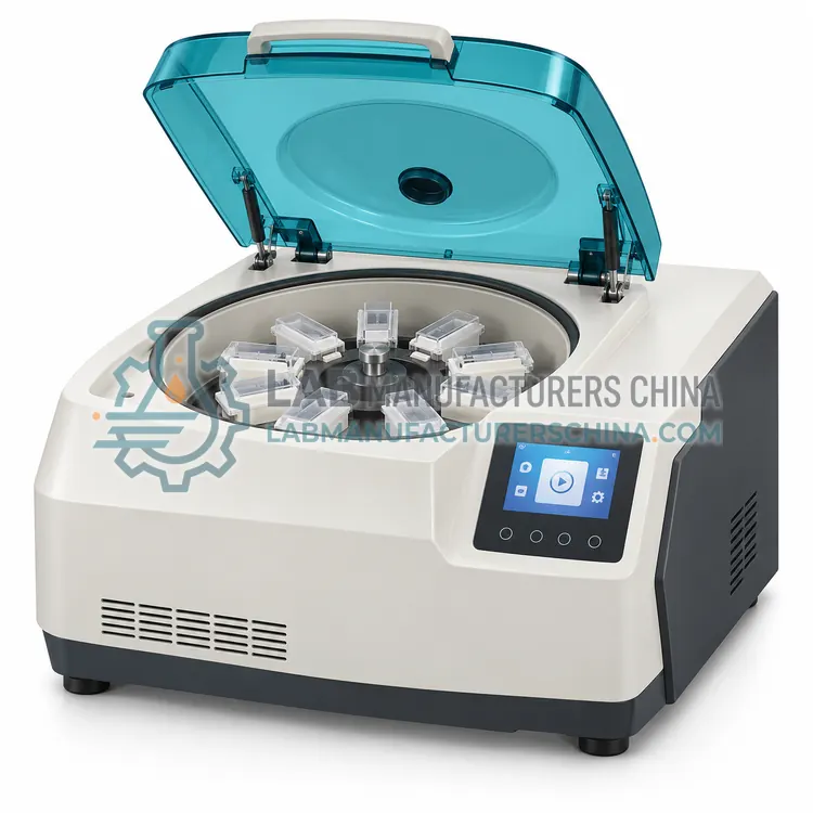 CCYE细胞学设备Cytology equipment covers the instruments that concentrate, prepare and stain individual cells for microscopic examination rather than intact tissue. It serves hospital cytology laboratories, cervical screening programmes and research facilities that examine exfoliated cells, body fluids and fine-needle aspirates.
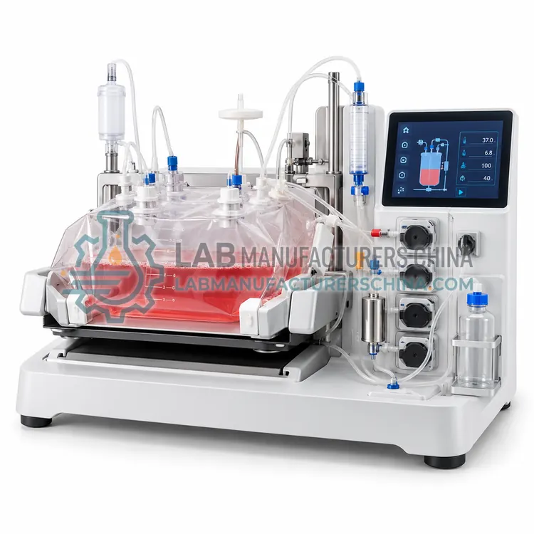 CCLE临床实验室设备Clinical laboratory equipment covers the instruments that test patient specimens such as blood, urine and body fluids to support diagnosis and monitoring. It includes clinical analyzers, sample processing devices, centrifuges, microscopes and laboratory automation used in hospital, reference and clinic laboratories.
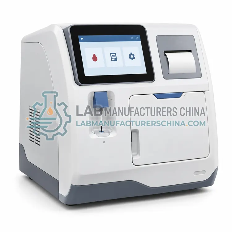 CDLE诊断实验室设备Diagnostic laboratory equipment covers instruments used to detect disease markers, pathogens and genetic targets in patient specimens. It includes diagnostic analyzers, immunodiagnostic and molecular diagnostic systems, point-of-care devices and sample preparation equipment used in hospitals, reference laboratories, clinics and public health settings.
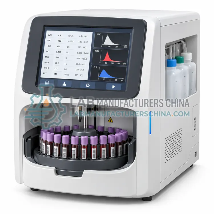 CHEA血液分析仪Hematology analyzers are instruments that count and characterize blood cells in whole blood or body fluids. They measure red cells, white cells, platelets and hemoglobin by impedance, light scatter or fluorescence, and are used by hospital laboratories, clinics and blood centers for complete blood counts and cell differentials.