
Urinalysis Analyzers
尿液分析仪
CURA · Clinical & Diagnostic · 3 product types · 15 models
By Lab Manufacturers China Technical TeamLast reviewed
What are Urinalysis Analyzers?
Urinalysis analyzers measure chemical and microscopic properties of urine to screen for renal, urinary tract and metabolic disease. They read reagent strips photometrically or classify particles by flow cytometry or imaging, and are used in hospital laboratories, clinics and screening programmes.
3 product types in Urinalysis Analyzers
The 3 product types below are grouped by the exact instrument you need.
- USR5 models
Urine Strip Readers
尿条读取仪
Urine strip readers measure the colour of reagent strip pads by reflectance photometry and convert it into semiquantitative results for glucose, protein, blood, leukocyte esterase, nitrite, ketones, bilirubin, urobilinogen, pH and specific gravity. They serve clinics, wards and screening programmes.
- AUS5 models
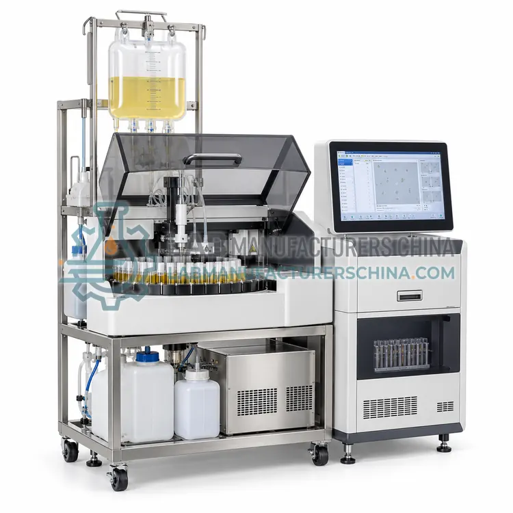
Automated Urinalysis Systems
全自动尿液分析系统
Automated urinalysis systems combine reagent strip chemistry with sediment analysis on one platform, so a single urine sample yields a complete report. They handle sample loading, mixing, measurement and reporting automatically and serve hospital laboratories with routine urinalysis workloads.
- USA5 models
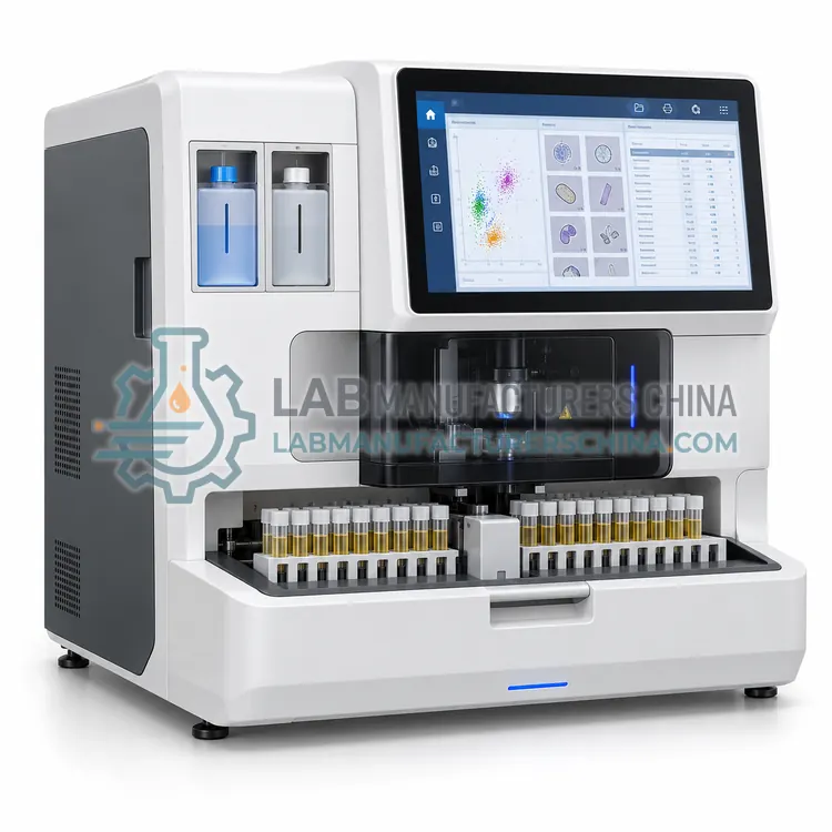
Urine Sediment Analyzers
尿沉渣分析仪
Urine sediment analyzers count and classify particles in urine sediment, such as red cells, white cells, epithelial cells, casts, crystals, bacteria and yeast. They replace part of manual microscopy using flow cytometry or digital imaging and are used in hospital and outpatient urinalysis workflows.

What are Urinalysis Analyzers?
Urinalysis analyzers automate the examination of urine, one of the most frequently requested laboratory tests. Chemical analysis uses reagent strips that react with glucose, protein, blood, leukocyte esterase, nitrite, ketones, bilirubin, urobilinogen, pH, specific gravity and sometimes ascorbate or microalbumin, and the strip reader measures the reflectance or absorbance of each pad. Microscopic analysis examines cells, casts, crystals, bacteria and yeast in the sediment, either by flow cytometry with scatter and fluorescence, or by digital imaging of a sediment slide, or by the classical centrifuge and microscope method. The category therefore covers strip readers, complete automated urinalysis systems that combine chemistry and sediment on one platform, and dedicated sediment analyzers. Laboratories select equipment according to daily specimen volume, whether sediment results are required, the particle classification the clinicians need, and the level of automation, connectivity and cross-contamination control required.
How do Urinalysis Analyzers work?
Reagent strip analyzers press the strip against a light source and read the colour of each pad with photodiodes or a camera at defined wavelengths. The colour change is converted to a semiquantitative result, usually on a scale of negative, trace, 1+, 2+, 3+ and 4+, or to a numeric value for pH and specific gravity. Reflectance photometry compares the light reflected from the reacted pad with a reference, and compensation channels correct for urine colour and for ascorbic acid interference. Flow cytometry sediment analyzers dilute urine, stain it with a fluorescent dye that labels nucleic acids and cell membranes, and pass it through a flow cell where forward scatter, side scatter and fluorescence classify each particle by size, granularity and stain uptake. Digital imaging analyzers centrifuge or allow sedimentation in a chamber and photograph the sediment, then classify particles with image recognition software. Automated systems transfer the same specimen to both chemistry and sediment modules.
Where are Urinalysis Analyzers used?
Urinalysis supports the detection and monitoring of urinary tract infection, kidney disease, diabetes, liver and biliary disorders, haematuria and kidney stones. Screening programmes use urine chemistry for antenatal care, pre-employment and insurance examinations, and school health checks. Hospital laboratories combine chemical and microscopic analysis for nephrology, urology and obstetric wards, and follow patients after transplantation or during chemotherapy. Specific gravity and pH results guide fluid status and stone prevention advice. Microalbumin measurement on strips or analyzers supports diabetic nephropathy screening. Sediment examination helps distinguish glomerular from non-glomerular bleeding and identifies casts, crystals and dysmorphic red cells. Veterinary laboratories use similar methods for companion animals, and urine drug screening uses separate immunoassay strips.
Which standards apply to Urinalysis Analyzers?
Urinalysis in medical laboratories follows ISO 15189, with method verification and precision studies guided by CLSI GP16 for urinalysis and CLSI EP05 and EP15. Reagent strip performance is evaluated against reference methods for each analyte, including the icteric, ketone, protein and glucose pads, and participant laboratories join external quality assessment schemes for urine chemistry and sediment. The Clinical and Laboratory Standards Institute publishes guidelines for urine sediment examination, and ISO 15197 applies where blood glucose rather than urine glucose is measured. Electrical safety follows IEC 61010-1 and IEC 61010-2-101. Market registration may require CE marking under the IVD Regulation, NMPA registration in China or FDA clearance in the United States, and reagent strip labelling follows ISO 18113-1.
How do I select the right urinalysis analyzers equipment?
Decide first whether the laboratory must report urine sediment, because that determines whether a strip reader is sufficient or an automated system is needed. Count daily specimens and consider peak morning workload, since urine samples cluster in the early shift. A clinic processing fewer than one hundred strips per day can use a bench-top or portable strip reader, while a hospital laboratory with several hundred specimens needs a strip reader with automatic strip feeding or a combined chemistry and sediment system. For sediment, compare flow cytometry, which gives counts of red cells, white cells, epithelial cells, casts and bacteria with low labour input, against digital imaging, which preserves an image for review. Check cross-contamination control, barcode reading, LIS connectivity, the report format the clinicians expect, and the supply and shelf life of strips, controls and calibrators.
Urinalysis Analyzers comparison
| Subcategory | Principle | Typical use | Key spec |
|---|---|---|---|
| Urine Strip Readers | Reflectance photometry of reagent strip pads at defined wavelengths | Chemical urine screening in clinics and low-volume laboratories | Parameters measured and strips per hour |
| Automated Urinalysis Systems | Combined strip chemistry and sediment analysis on one platform | Hospital routine urinalysis with walkaway operation | Throughput and combined module options |
| Urine Sediment Analyzers | Flow cytometry or digital imaging classification of urine particles | Microscopic screening and particle counting of urine sediment | Particle types reported and sample volume |
Frequently asked questions about Urinalysis Analyzers
A chemistry analyzer measures glucose, protein, blood, leukocyte esterase, nitrite, ketones, bilirubin, urobilinogen, pH and specific gravity from reagent strips, and some add microalbumin or creatinine. Sediment analyzers additionally count red cells, white cells, epithelial cells, casts, crystals and bacteria in the urine.
A strip reader covers chemical screening and suits clinics and low-volume sites. A full automated system is appropriate when sediment results are required routinely, when specimen numbers exceed a few hundred per day, or when standardised particle counts and reduced manual microscopy time are important.
Midstream clean-catch urine is preferred for culture and routine analysis, collected in a sterile container and analysed within two hours at room temperature. If delay is unavoidable, refrigerate at 2 to 8 degrees Celsius and allow the specimen to reach room temperature before analysis, because cells and casts degrade and crystals may precipitate.
Ascorbic acid can suppress glucose, blood and bilirubin pads, strong urinary colour interferes with reflectance reading, and high specific gravity reduces reactivity. Contamination from disinfectants and delay in transport can also alter results, so strip readers with colour and ascorbate compensation are preferred for routine work.
Sediment analyzers are calibrated by the manufacturer against reference microscopy, and laboratories verify agreement with manual microscopy on a defined number of samples across a range of particle concentrations. Daily quality control uses stabilised particle suspensions or commercial control material, and external quality assessment schemes compare particle counts with peer laboratories.
Related categories
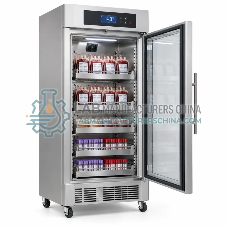 CBBE血库设备Blood bank equipment covers the refrigerators, plasma freezers, collection monitors, tube sealers and plasma thawers used to collect, process, store and issue blood components. It supports blood centers, hospital transfusion services and plasma facilities that must hold defined temperatures and keep traceable records.
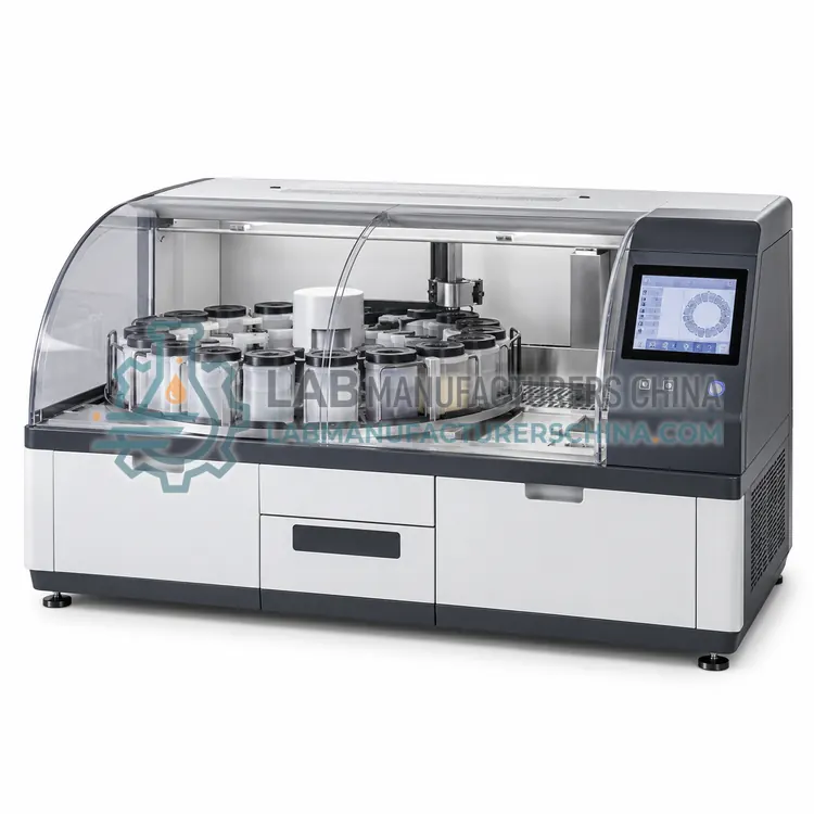 CPEQ病理设备Pathology equipment covers the instruments that turn surgical and biopsy specimens into stained, diagnosable slides. It includes grossing stations, tissue processors, embedding stations, microtomes and slide stainers, used in hospital pathology departments, reference laboratories, veterinary and research histology services.
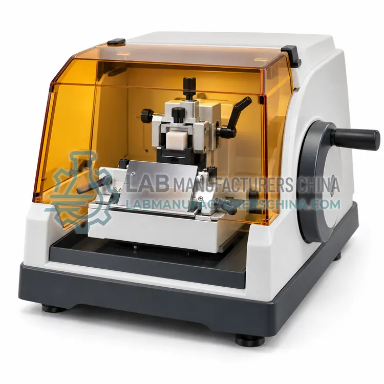 CHIE组织学设备Histology equipment covers the instruments used to embed, section, float, dry and stain tissue samples for microscopic examination. It serves hospital pathology laboratories, research histology facilities and contract research organizations that turn biopsy and necropsy specimens into stained slides for diagnosis and study.
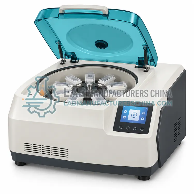 CCYE细胞学设备Cytology equipment covers the instruments that concentrate, prepare and stain individual cells for microscopic examination rather than intact tissue. It serves hospital cytology laboratories, cervical screening programmes and research facilities that examine exfoliated cells, body fluids and fine-needle aspirates.