Lab Manufacturers China

Urine Sediment Analyzers

尿沉渣分析仪

USA · Urinalysis Analyzers · 5 models

By Lab Manufacturers China Technical TeamLast reviewed Standards: ISO 15189, CLSI GP16, CLSI EP05

Standards referenced

  • ISO 15189
  • CLSI GP16
  • CLSI EP05
  • CLSI EP15
  • IEC 61010-1
  • IEC 61010-2-101

What are Urine Sediment Analyzers?

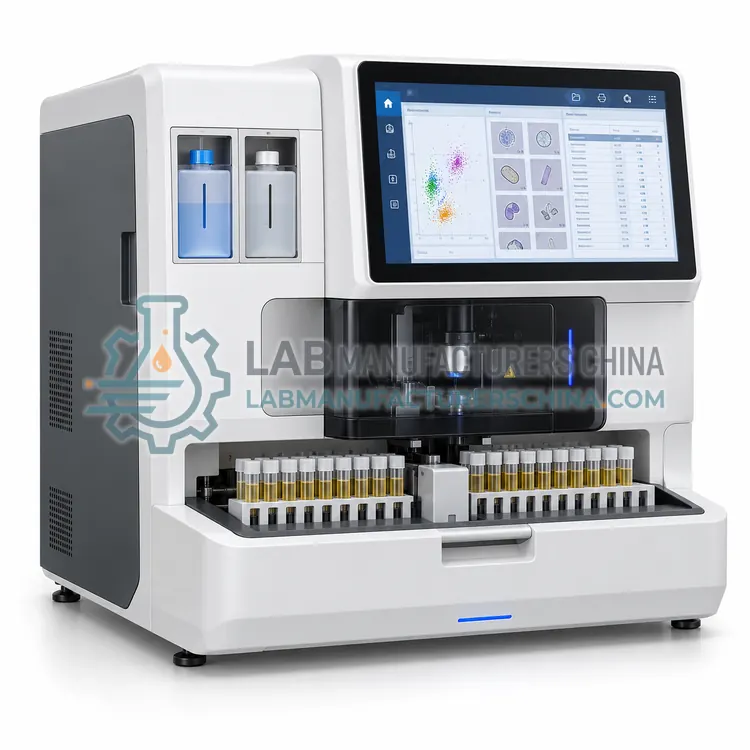
Urine sediment analyzers count and classify particles in urine sediment, such as red cells, white cells, epithelial cells, casts, crystals, bacteria and yeast. They replace part of manual microscopy using flow cytometry or digital imaging and are used in hospital and outpatient urinalysis workflows.

5 models of Urine Sediment Analyzers

The 5 models below are current configurations; specifications are confirmed on quotation.

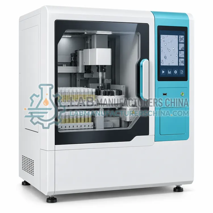
Flow Cytometry Urine Sediment Analyzer

流式细胞术尿沉渣分析仪

  • USA-0001
  • Urine Sediment Analyzers
  • CE (to be confirmed)
  • ISO 13485 manufacturing (to be confirmed)
  • NMPA class II registration (to be confirmed)
Detection principle:
Flow cytometry with nucleic acid fluorescence staining
Particle classes:
RBC, WBC, epithelial cells, casts, crystals, bacteria, yeast
Throughput:
50–100 samples/h
Sample volume:
0.5–1.0 mL per analysis
Reporting units:
Counts per microlitre with high-power field equivalents
Reagents:
Sheath fluid, diluent and fluorescent stain

A flow cytometry urine sediment analyzer stains diluted urine with a fluorescent nucleic acid dye and classifies particles by scatter and fluorescence signals, reporting counts per microlitre. It suits laboratories that need rapid, standardised sediment counts with minimal manual preparation.

Request a quote
View details

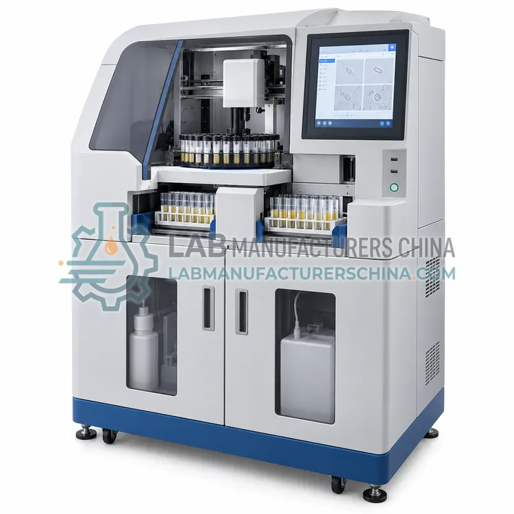
Digital Imaging Urine Sediment Analyzer

数字成像尿沉渣分析仪

  • USA-0002
  • Urine Sediment Analyzers
  • CE (to be confirmed)
  • ISO 13485 manufacturing (to be confirmed)
  • NMPA class II registration (to be confirmed)
Imaging method:
Camera imaging in a defined-depth chamber
Particle classes:
RBC, WBC, epithelial cells, casts, crystals, bacteria, yeast
Throughput:
40–80 samples/h
Sample volume:
0.5–1.5 mL
Images per sample:
Up to 500 images stored
Resolution:
Approximately 1 µm per pixel

A digital imaging urine sediment analyzer photographs particles in a thin counting chamber and classifies them by pattern recognition, storing images with each result. It suits laboratories that want visual evidence and on-screen verification of sediment findings.

Request a quote
View details

Automated Urine Particle Analyzer

尿液颗粒自动分析仪

  • USA-0003
  • Urine Sediment Analyzers
  • CE (to be confirmed)
  • ISO 13485 manufacturing (to be confirmed)
  • NMPA class II registration (to be confirmed)
Detection principle:
Flow cytometry or impedance with light scatter
Reported particles:
RBC, WBC, epithelial cells, bacteria, casts, crystals, yeast
Throughput:
60–100 samples/h
Sample volume:
0.5–1.0 mL uncentrifuged urine
Reporting units:
Cells per microlitre with field equivalents
Flags:
Bacterial overgrowth, atypical particle pattern

An automated urine particle analyzer is optimised for counting and differentiating particles in uncentrifuged urine with minimal sample preparation, reporting red cells, white cells, epithelial cells, bacteria and casts. It suits laboratories screening large numbers of urine samples for infection and renal disease.

Request a quote
View details

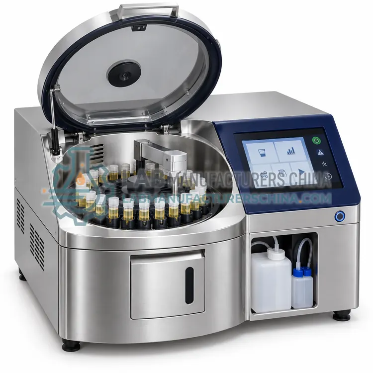
Urine Sediment Analyzer with Integrated Centrifugation

带集成离心功能的尿沉渣分析仪

  • USA-0004
  • Urine Sediment Analyzers
  • CE (to be confirmed)
  • ISO 13485 manufacturing (to be confirmed)
  • NMPA class II registration (to be confirmed)
Preparation:
Automated centrifugation and resuspension
Centrifugation force:
400–1500 × g adjustable
Sample volume:
5–15 mL urine
Concentration factor:
10–50 fold depending on protocol
Detection:
Imaging or flow counting of concentrated sediment
Throughput:
15–40 samples/h

This analyzer centrifuges the urine, resuspends the sediment and images or counts it in a concentrated form, which increases sensitivity at low particle counts. It suits laboratories that need improved detection of low-level haematuria or cellular sediment.

Request a quote
View details

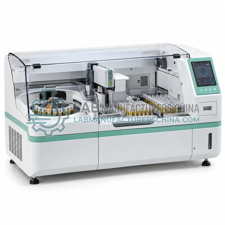
Benchtop Urine Sediment Analyzer with Autosampler

带自动进样器的台式尿沉渣分析仪

  • USA-0005
  • Urine Sediment Analyzers
  • CE (to be confirmed)
  • ISO 13485 manufacturing (to be confirmed)
  • NMPA class II registration (to be confirmed)
Autosampler capacity:
40–80 tubes with barcode identification
Throughput:
60–120 samples/h
Detection principle:
Flow cytometry or digital imaging
Sample volume:
0.5–1.5 mL per sample
Carryover:
< 1 % for red and white cell counts
Reported particles:
RBC, WBC, epithelial cells, casts, crystals, bacteria, yeast

This benchtop analyzer adds a sample carousel to the sediment analysis module, so a batch of urine tubes is processed without individual handling. It suits mid-size laboratories that need sediment counts on hundreds of samples per day in a compact footprint.

Request a quote
View details

Compare Urine Sediment Analyzers models

ModelDetection principleParticle classesThroughputSample volumeReporting units
Flow Cytometry Urine Sediment AnalyzerUSA-0001Flow cytometry with nucleic acid fluorescence stainingRBC, WBC, epithelial cells, casts, crystals, bacteria, yeast50–100 samples/h0.5–1.0 mL per analysisCounts per microlitre with high-power field equivalents
Digital Imaging Urine Sediment AnalyzerUSA-0002—RBC, WBC, epithelial cells, casts, crystals, bacteria, yeast40–80 samples/h0.5–1.5 mL—
Automated Urine Particle AnalyzerUSA-0003Flow cytometry or impedance with light scatter—60–100 samples/h0.5–1.0 mL uncentrifuged urineCells per microlitre with field equivalents
Urine Sediment Analyzer with Integrated CentrifugationUSA-0004——15–40 samples/h5–15 mL urine—
Benchtop Urine Sediment Analyzer with AutosamplerUSA-0005Flow cytometry or digital imaging—60–120 samples/h0.5–1.5 mL per sample—

Indicative values for this product class. Final specifications are confirmed on the quotation and in the calibration documents supplied with the instrument.

How do Urine Sediment Analyzers work?

Urine sediment contains formed elements that indicate renal or urinary tract disease. Automated analyzers detect them in one of two ways. Flow cytometry dilutes urine, stains nucleic acids and cell membranes with a fluorescent dye, and passes the particles through a flow cell where forward scatter indicates size, side scatter indicates granularity and fluorescence indicates nucleic acid content; clustering software classifies the particles and reports counts per volume. Digital imaging introduces a diluted sample into a thin chamber, photographs the particles and applies pattern recognition to classify them, storing images for review. Either method reports counts per microlitre or per high-power field, together with flags when the result requires confirmation. Some instruments include a centrifugation step, and all of them depend on adequate sample mixing, correct dilution and clean optical or fluidic paths.

Which Urine Sediment Analyzers variants are available?

  • Flow Cytometry Urine Sediment Analyzer — A flow cytometry urine sediment analyzer stains diluted urine with a fluorescent nucleic acid dye and classifies particles by scatter and fluorescence signals, reporting counts per microlitre.
  • Digital Imaging Urine Sediment Analyzer — A digital imaging urine sediment analyzer photographs particles in a thin counting chamber and classifies them by pattern recognition, storing images with each result.
  • Automated Urine Particle Analyzer — An automated urine particle analyzer is optimised for counting and differentiating particles in uncentrifuged urine with minimal sample preparation, reporting red cells, white cells, epithelial cells, bacteria and casts.
  • Urine Sediment Analyzer with Integrated Centrifugation — This analyzer centrifuges the urine, resuspends the sediment and images or counts it in a concentrated form, which increases sensitivity at low particle counts.
  • Benchtop Urine Sediment Analyzer with Autosampler — This benchtop analyzer adds a sample carousel to the sediment analysis module, so a batch of urine tubes is processed without individual handling.

How do I choose the right variant?

Choose the detection method according to what your clinicians need. Flow cytometry gives fast counts of the main particle classes with minimal labour but limited morphological detail, while digital imaging provides images and better distinction between similar particles such as casts and crystals. Decide whether the analyzer must work with urine that has not been centrifuged, because some instruments require a pre-centrifuged or specially prepared sample. Check the reported particle classes, the counting units, the sample volume and the review rules, and confirm that the instrument can be integrated with the chemistry analyzer so that results appear in one report. Plan for periodic comparison with manual microscopy, and for external quality assessment covering urine sediment.

Where are Urine Sediment Analyzers used?

Urine Sediment Analyzers are specified in urinary tract infection assessment, glomerular and non-glomerular haematuria differentiation, renal disease monitoring and transplant follow-up and urology and nephrology outpatient testing.

Which standards and certifications apply?

Work with Urine Sediment Analyzers is commonly governed by ISO 15189, CLSI GP16, CLSI EP05, CLSI EP15, IEC 61010-1 and IEC 61010-2-101. Instrument certifications such as CE marking are confirmed per configuration and never assumed.

What are the key considerations before purchase?

Verify performance at installation by comparing results with manual microscopy on a defined number of samples spanning low, normal and high particle counts, and document the agreement, since counts per volume and per high-power field are not directly interchangeable. Run control material or stabilised particle suspensions at the recommended frequency, and clean the flow cell or imaging chamber as instructed, because deposits create artefacts or block the sample path. Maintain the sheath fluid supply where used, and check that it is within its expiry date. Define and document review rules, train staff to interpret the images or counts, and keep records linking stored images to patient results for the retention period required locally.

Frequently asked questions about Urine Sediment Analyzers

Typical reports include red blood cells, white blood cells, epithelial cells, casts, crystals, bacteria and yeast, and some instruments add squamous epithelial cells, hyaline casts or pathological casts separately, together with a flag when the pattern suggests infection or renal disease.

Need help specifying Urine Sediment Analyzers?

Request a quote