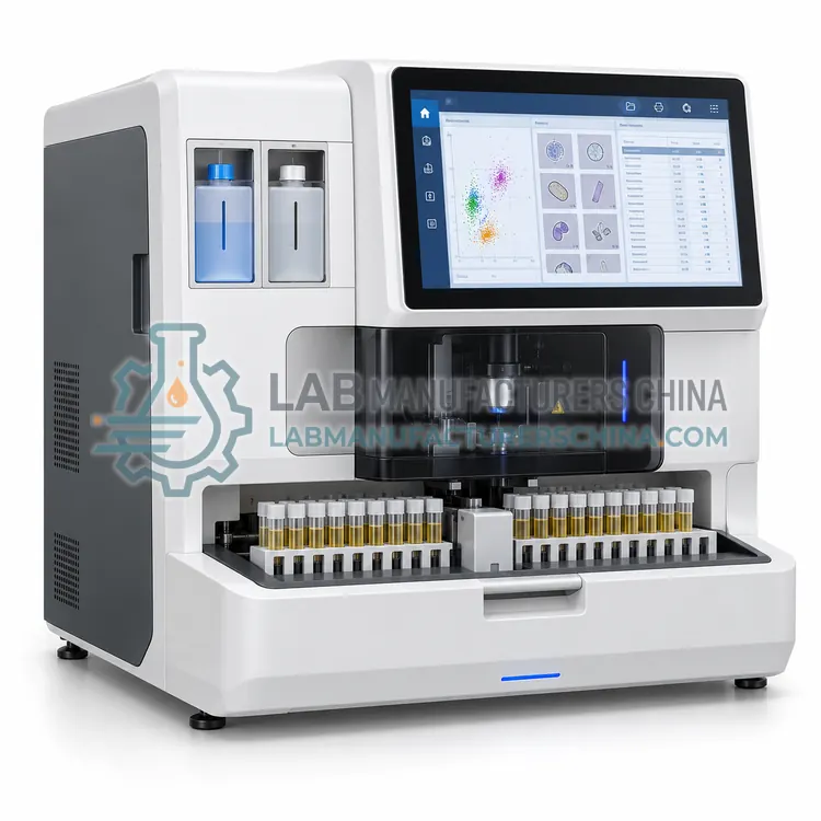
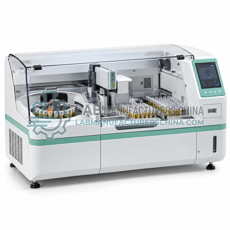
Urine Sediment Analyzer with Integrated Centrifugation
带集成离心功能的尿沉渣分析仪
- SKU USA-0004
- Urine Sediment Analyzers
- CE (to be confirmed)
- ISO 13485 manufacturing (to be confirmed)
- NMPA class II registration (to be confirmed)
Price on request — FOB / CIF quoted per order
Prices depend on configuration, quantity and destination port.
Key specifications
| Preparation | Automated centrifugation and resuspension |
|---|---|
| Centrifugation force | 400–1500 × g adjustable |
| Sample volume | 5–15 mL urine |
| Concentration factor | 10–50 fold depending on protocol |
| Detection | Imaging or flow counting of concentrated sediment |
| Throughput | 15–40 samples/h |
Request a quote Download brochure · to be provided
By Lab Manufacturers China Technical TeamLast reviewed Standards: ISO 15189, CLSI GP16, CLSI EP05
What is the Urine Sediment Analyzer with Integrated Centrifugation?
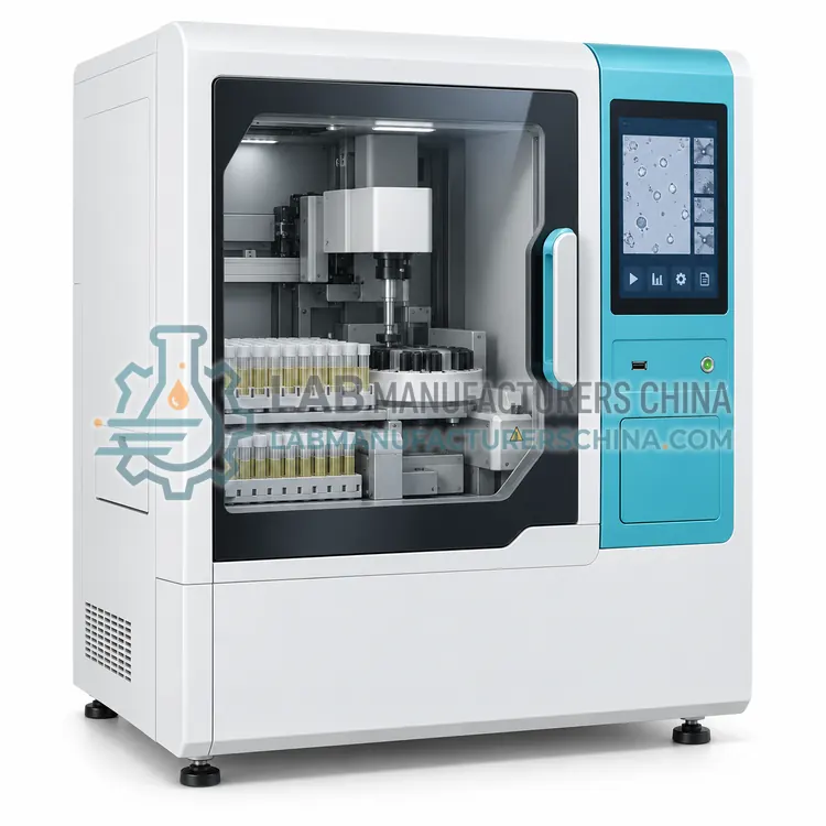
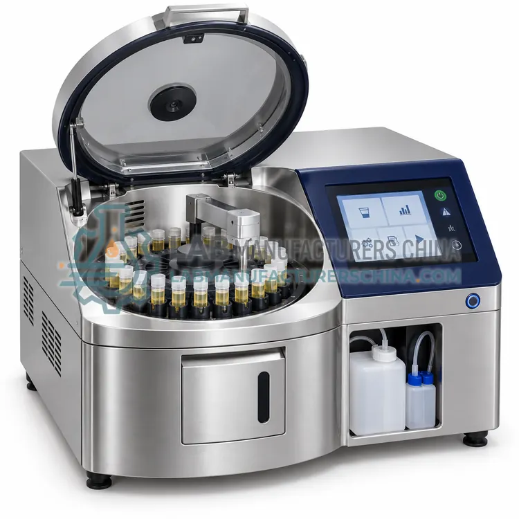
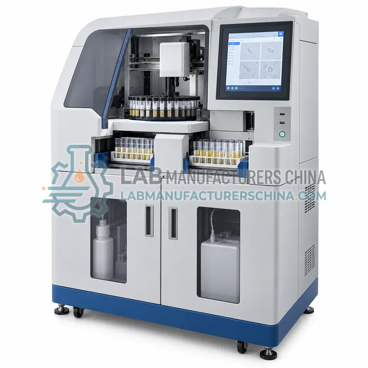
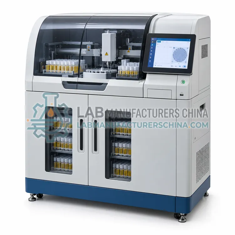
This analyzer centrifuges the urine, resuspends the sediment and images or counts it in a concentrated form, which increases sensitivity at low particle counts. It suits laboratories that need improved detection of low-level haematuria or cellular sediment.
What is the Urine Sediment Analyzer with Integrated Centrifugation?
This analyzer centrifuges the urine, resuspends the sediment and images or counts it in a concentrated form, which increases sensitivity at low particle counts. It suits laboratories that need improved detection of low-level haematuria or cellular sediment.
Overview
The instrument automates the classical preparation: a measured urine volume is centrifuged at a defined relative centrifugal force and time, supernatant is removed, and the sediment is resuspended in a small volume before being applied to a counting chamber or imaged directly. Concentrating the sediment lowers the detection limit, so low numbers of red cells, white cells or casts can be detected that might fall below the reporting threshold of direct methods. The procedure requires careful control of centrifugation force, decanting and resuspension, and the software records these parameters with the result. Sample volume is larger than for direct methods, typically 5 to 15 millilitres, and throughput is lower, so the approach suits targeted rather than screening workloads.
How does it work?
Urine sediment contains formed elements that indicate renal or urinary tract disease. Automated analyzers detect them in one of two ways. Flow cytometry dilutes urine, stains nucleic acids and cell membranes with a fluorescent dye, and passes the particles through a flow cell where forward scatter indicates size, side scatter indicates granularity and fluorescence indicates nucleic acid content; clustering software classifies the particles and reports counts per volume. Digital imaging introduces a diluted sample into a thin chamber, photographs the particles and applies pattern recognition to classify them, storing images for review. Either method reports counts per microlitre or per high-power field, together with flags when the result requires confirmation. Some instruments include a centrifugation step, and all of them depend on adequate sample mixing, correct dilution and clean optical or fluidic paths.
What are the key features?
- Automated centrifugation with controlled force and time
- Supernatant removal and controlled sediment resuspension
- Concentrated sediment imaging or counting chamber analysis
- Enhanced detection of low particle counts
- Sample and preparation parameters recorded with results
- Larger sample volume suited to targeted testing
What are the technical specifications?
A typical urine sediment analyzer with integrated centrifugation configuration offers preparation of Automated centrifugation and resuspension, centrifugation force of 400–1500 × g adjustable, sample volume of 5–15 mL urine, concentration factor of 10–50 fold depending on protocol, detection of Imaging or flow counting of concentrated sediment and throughput of 15–40 samples/h. Values are indicative of the product class; the exact figures for each configuration are confirmed on the quotation and the calibration documents supplied with the instrument.
Which applications is it used for?
The Urine Sediment Analyzer with Integrated Centrifugation is used in investigation of low-level haematuria, renal biopsy follow-up with minimal sediment, urology clinics assessing haematuria and research studies requiring concentrated sediment. Across the wider Urine Sediment Analyzers class, laboratories also deploy it for urinary tract infection assessment, glomerular and non-glomerular haematuria differentiation and renal disease monitoring and transplant follow-up.
Which standards and certifications apply?
Relevant standards and methods include ISO 15189, CLSI GP16, CLSI EP05, CLSI EP15, IEC 61010-1 and IEC 61010-2-101. Certification candidates for this model are CE, ISO 13485 manufacturing and NMPA class II registration; each is to be confirmed for the specific configuration before shipment, and certificates are supplied only where they have been issued.
Verify performance at installation by comparing results with manual microscopy on a defined number of samples spanning low, normal and high particle counts, and document the agreement, since counts per volume and per high-power field are not directly interchangeable. Run control material or stabilised particle suspensions at the recommended frequency, and clean the flow cell or imaging chamber as instructed, because deposits create artefacts or block the sample path. Maintain the sheath fluid supply where used, and check that it is within its expiry date. Define and document review rules, train staff to interpret the images or counts, and keep records linking stored images to patient results for the retention period required locally.
How do I select the right Urine Sediment Analyzer with Integrated Centrifugation?
Specify this variant when sensitivity at low particle counts is the priority, or when the laboratory currently performs a full manual centrifugation protocol and wants to standardise it. In the RFQ confirm the centrifugation parameters, the concentration factor, the sample volume required and how preparation parameters are recorded for traceability.
How does it compare with other Urine Sediment Analyzers variants?
Choose the detection method according to what your clinicians need. Flow cytometry gives fast counts of the main particle classes with minimal labour but limited morphological detail, while digital imaging provides images and better distinction between similar particles such as casts and crystals. Decide whether the analyzer must work with urine that has not been centrifuged, because some instruments require a pre-centrifuged or specially prepared sample. Check the reported particle classes, the counting units, the sample volume and the review rules, and confirm that the instrument can be integrated with the chemistry analyzer so that results appear in one report. Plan for periodic comparison with manual microscopy, and for external quality assessment covering urine sediment.
Full specifications
| Preparation | Automated centrifugation and resuspension |
|---|---|
| Centrifugation force | 400–1500 × g adjustable |
| Sample volume | 5–15 mL urine |
| Concentration factor | 10–50 fold depending on protocol |
| Detection | Imaging or flow counting of concentrated sediment |
| Throughput | 15–40 samples/h |
| Reported particles | RBC, WBC, epithelial cells, casts, crystals, bacteria |
| Data storage | 5,000 results with preparation parameters |
| Interfaces | LAN with HL7 or ASTM, USB |
Indicative values for this product class. Final specifications are confirmed on the quotation and in the calibration documents supplied with the instrument.
Applications
- Investigation of low-level haematuria
- Renal biopsy follow-up with minimal sediment
- Urology clinics assessing haematuria
- Research studies requiring concentrated sediment
Key features
- Automated centrifugation with controlled force and time
- Supernatant removal and controlled sediment resuspension
- Concentrated sediment imaging or counting chamber analysis
- Enhanced detection of low particle counts
- Sample and preparation parameters recorded with results
- Larger sample volume suited to targeted testing
Frequently asked questions about Urine Sediment Analyzer with Integrated Centrifugation
Concentrating the sediment from a larger urine volume increases the number of particles in the analysed chamber, so low counts that would be below the detection limit of a direct method become measurable. The trade-off is a longer preparation and more manual handling of the sample.
Relative centrifugal force and time must match the validated protocol, since differences change recovery of cells and casts. Many instruments monitor rotor speed and log the parameters with the result, which is useful when a result is questioned or when comparing platforms.
It can, but throughput of 15 to 40 samples per hour makes it less suitable than direct analyzers for large screening workloads. Laboratories typically use it for targeted investigations or as a reference method alongside a faster screening instrument.
Typically 5 to 15 millilitres, which is more than direct methods require, so the urine must be collected in a suitable container and analysed within the stability window. Inadequate volume reduces the concentration factor and hence the sensitivity gain.
Typical reports include red blood cells, white blood cells, epithelial cells, casts, crystals, bacteria and yeast, and some instruments add squamous epithelial cells, hyaline casts or pathological casts separately, together with a flag when the pattern suggests infection or renal disease.
Agreement is good for red cells, white cells and epithelial cells in the normal to high range, but casts and crystals are harder to classify automatically, and low counts near the detection limit are less precise. Laboratories must verify agreement locally and define when microscopy is required.
Yes. Microscope examination is required when automated results are abnormal, when the clinical picture suggests a discrepancy, when atypical or unclassified particles appear, and when a specific morphology such as dysmorphic red cells is requested. Most laboratories keep microscopy for these reflex situations.