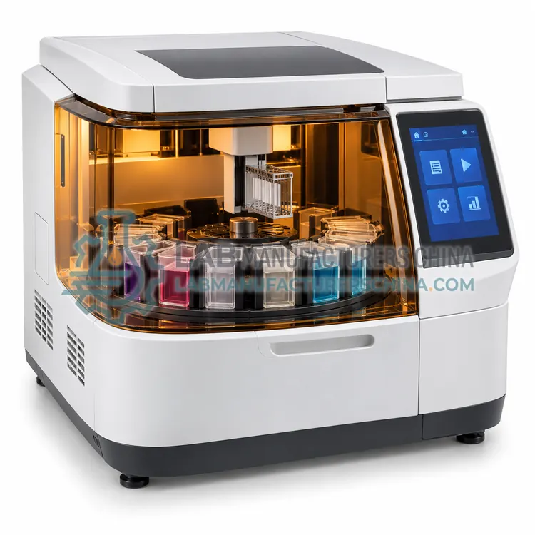
How do Histology Stainers work?
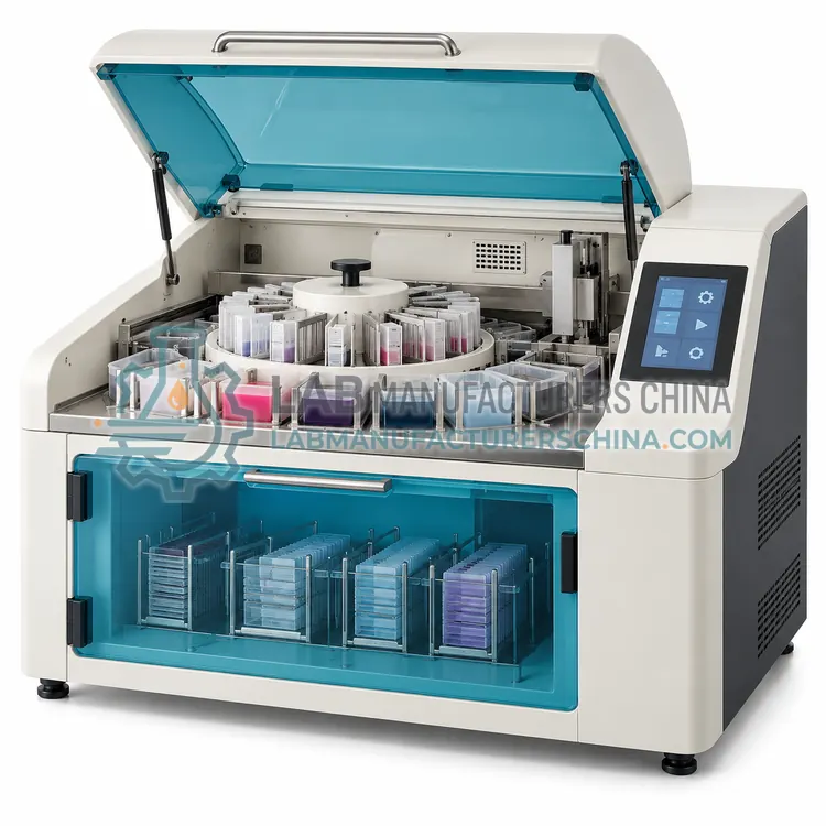
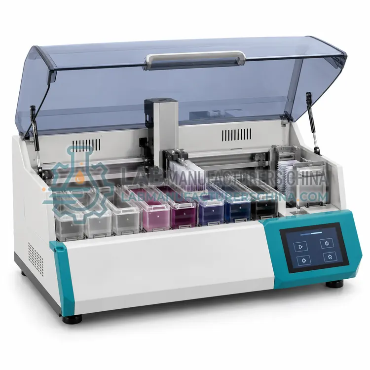
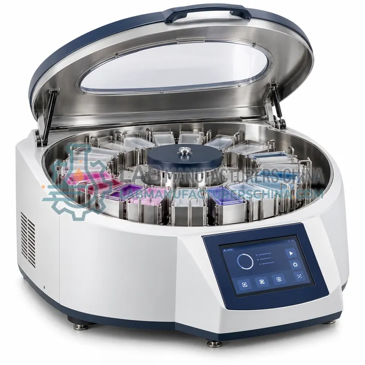
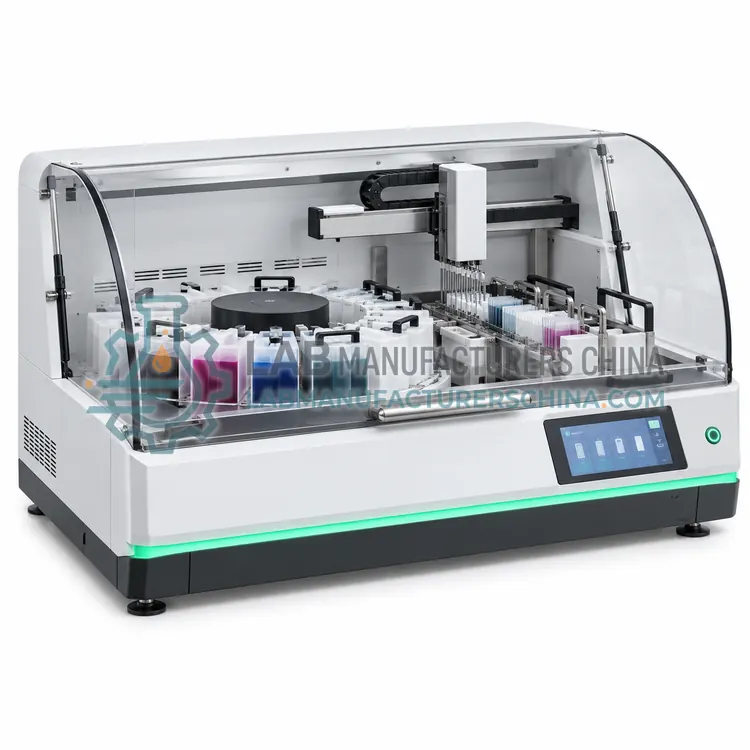
A histology stainer automates the sequence of deparaffinisation, hydration, staining, differentiation, dehydration and clearing that a technician would perform by hand. Slides are held in racks or on a conveyor, and the instrument moves them between reagent baths for set dwell times, then transfers them to running water or a rinse station. Hematoxylin stains nucleic acids blue, eosin counterstains cytoplasm pink, and differentiation in acid alcohol tunes the contrast. Special stains such as Periodic Acid-Schiff, Masson trichrome or reticulin use different reagent sequences. Consistency comes from controlled dwell time, reagent concentration, agitation and temperature, while carryover is managed by draining and rinse stations. Automated coverslipping may follow on the same platform.
Which Histology Stainers variants are available?
- Manual Histology Staining Station — A manual histology staining station groups staining baths, a running-water sink and a fume hood into one bench arrangement.
- Linear Slide Stainer — A linear slide stainer moves racks along a track through fixed reagent baths at set intervals, giving reproducible routine staining at moderate throughput.
- Automated H&E Slide Stainer — An automated H&E slide stainer programs deparaffinisation, staining, differentiation, dehydration and clearing as a single walk-away cycle.
- Automated Special Stains Slide Stainer — An automated special stains slide stainer runs protocols such as PAS, trichrome, reticulin or Gram stains with individually timed reagent steps.
- Compact Benchtop Slide Stainer — A compact benchtop slide stainer automates routine staining in a small footprint, typically handling 20 to 60 slides per run.
How do I choose the right variant?
Select a stainer by throughput, reagent menu and the level of walk-away automation required. Manual staining stations cost least and give full control for low volumes and teaching. Linear stainers move racks along a track through fixed baths and suit routine H&E at moderate volume. Fully automated H&E stainers program time, temperature and agitation, and can process hundreds of slides per hour. Special stain stainers handle individual protocols for trichrome, PAS, reticulin or Gram staining. Compact benchtop models fit small laboratories. Compare slides per run, reagent station count and volume, program capacity, carryover prevention and whether a coverslipper can be integrated. Confirm reagent compatibility and the cost of consumables.
Where are Histology Stainers used?
Histology Stainers are specified in routine H&E staining in pathology, special stains, immunohistochemistry counterstaining, research histology, pharmaceutical toxicology and cytology slide staining.
Which standards and certifications apply?
Work with Histology Stainers is commonly governed by IEC 61010-1, IEC 61010-2-101, IEC 61326-1 and ISO 15189. Instrument certifications such as CE marking are confirmed per configuration and never assumed.
What are the key considerations before purchase?
Install the stainer near a water supply and drain if it needs running water, and on a level bench away from direct sunlight that fades dyes. Reagents degrade with use and evaporation, so set replacement schedules based on slide counts and monitor staining quality with control slides. Clean baths and tubing regularly to prevent precipitate and microbial growth, and check that the differentiation step gives reproducible contrast. Record reagent changes, quality control results and maintenance. Train staff on loading orientation, since misplaced slides cause uneven staining, and on safe handling of xylene, alcohol and carcinogenic dyes.