Lab Manufacturers China

Cell Imaging

细胞成像系统

CEI · Cell Biology Equipment · 5 models

By Lab Manufacturers China Technical TeamLast reviewed Standards: IEC 60825-1, IEC 61010-1, IEC 61010-2-010

Standards referenced

  • IEC 60825-1
  • IEC 61010-1
  • IEC 61010-2-010
  • ISO 17025
  • ISO 20387

What are Cell Imaging?

Cell imaging systems visualise living and fixed cells with transmitted light and fluorescence, from routine inverted microscopes to confocal, live-cell and high-content platforms. They are used to assess morphology, protein localisation and dynamic processes in cell biology and drug discovery.

5 models of Cell Imaging

The 5 models below are current configurations; specifications are confirmed on quotation.

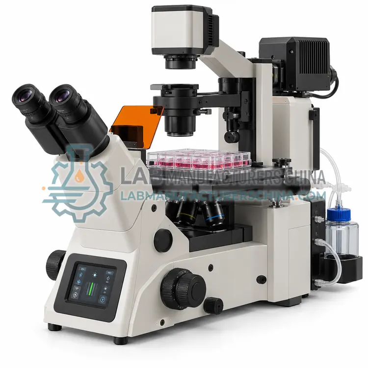
Inverted Fluorescence Microscope for Cell Culture

细胞培养倒置荧光显微镜

  • CEI-0001
  • Cell Imaging
  • CE (to be confirmed)
  • ISO 9001 manufacturing (to be confirmed)
  • IEC 61010-1 safety (to be confirmed)
Objectives:
4x, 10x, 20x, 40x and 60x options
Fluorescence channels:
2–5 filter cubes (DAPI, FITC, TRITC, Cy5)
Light source:
LED, 365/470/550/620 nm options
Camera:
sCMOS or CCD, 4–20 MP
Contrast methods:
Brightfield, phase contrast and relief contrast
Stage:
Fixed or motorised with multi-well plate insert

An inverted fluorescence microscope views cells from below through the base of a culture vessel, with LED excitation and two to five filter sets for routine morphology and immunofluorescence. It is the standard bench instrument in cell culture laboratories.

Request a quote
View details

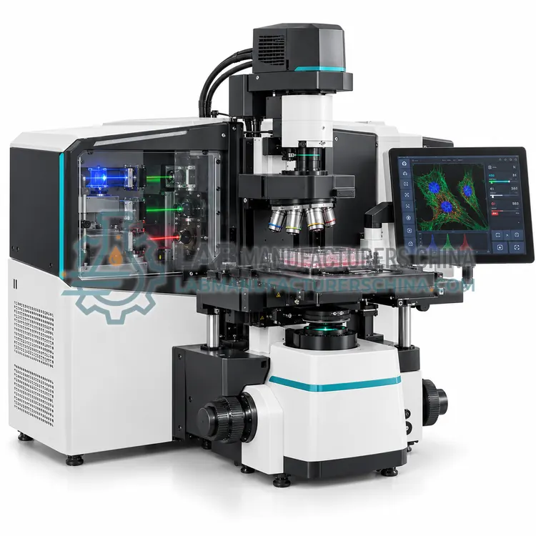
Confocal Laser Scanning Microscope

激光共聚焦显微镜

  • CEI-0002
  • Cell Imaging
  • CE (to be confirmed)
  • ISO 9001 manufacturing (to be confirmed)
  • IEC 61010-1 safety (to be confirmed)
Laser lines:
405, 488, 561 and 640 nm typical
Scanning:
Galvanometer mirrors, up to 512 x 512 at 4 fps
Detection:
Spectral detector or filter-based photomultipliers
Pinhole range:
1–5 Airy units adjustable
Lateral resolution:
approx. 200 nm lateral, 500 nm axial
Imaging depth:
approx. 50–150 µm in cleared or sparse tissue

A confocal laser scanning microscope uses a pinhole and scanning laser beams to reject out-of-focus light, producing optical sections through thick cells and tissue and enabling high-contrast three-dimensional reconstruction of subcellular structures.

Request a quote
View details

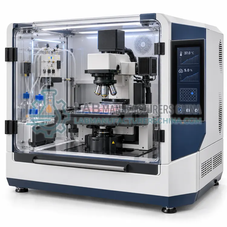
Live-Cell Imaging Incubation System

活细胞成像孵育系统

  • CEI-0003
  • Cell Imaging
  • CE (to be confirmed)
  • ISO 9001 manufacturing (to be confirmed)
  • IEC 61010-1 safety (to be confirmed)
Temperature control:
ambient to 45 °C, within ±0.2 °C
CO2 control:
0–10%, within ±0.1%
Humidity:
Maintained above 90% relative humidity
Oxygen control:
Optional 1–21%
Vessel compatibility:
Multi-well plates, dishes, flasks and chamber slides
Autofocus:
Hardware or image-based, continuous during acquisition

A live-cell imaging incubation system maintains temperature, carbon dioxide, humidity and often oxygen on the microscope stage while the software captures time-lapse sequences, so cell behaviour can be recorded continuously for hours or days.

Request a quote
View details

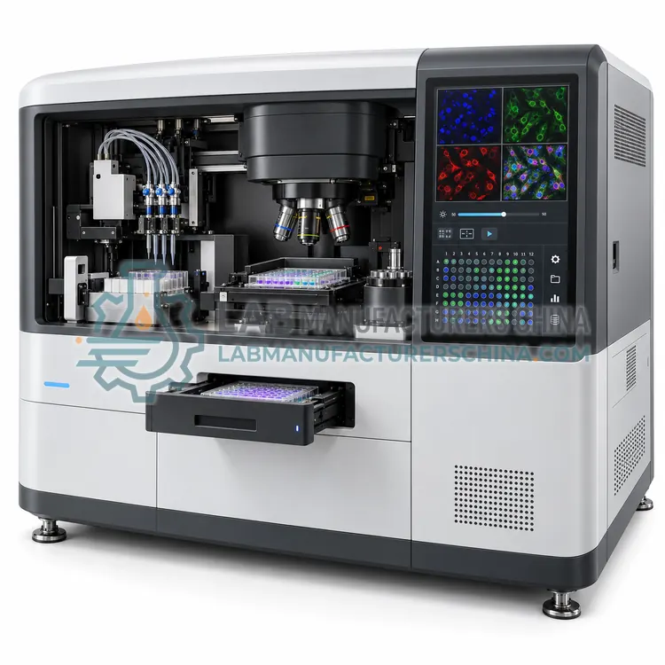
High-Content Screening Imaging System

高内涵筛选成像系统

  • CEI-0004
  • Cell Imaging
  • CE (to be confirmed)
  • ISO 9001 manufacturing (to be confirmed)
  • IEC 61010-1 safety (to be confirmed)
Plate formats:
6 to 1,536-well plates and flasks
Imaging channels:
4–8 fluorescence plus brightfield
Light sources:
LED or laser, 365–740 nm options
Objective set:
2x to 60x with autofocus
Throughput:
thousands to tens of thousands of images per day
Autofocus:
Image-based or hardware, per well

A high-content screening imaging system automates plate handling, autofocus, multi-channel image acquisition and analysis, extracting quantitative single-cell features from thousands of wells for compound screening, dose-response studies and phenotypic assay development in drug discovery.

Request a quote
View details

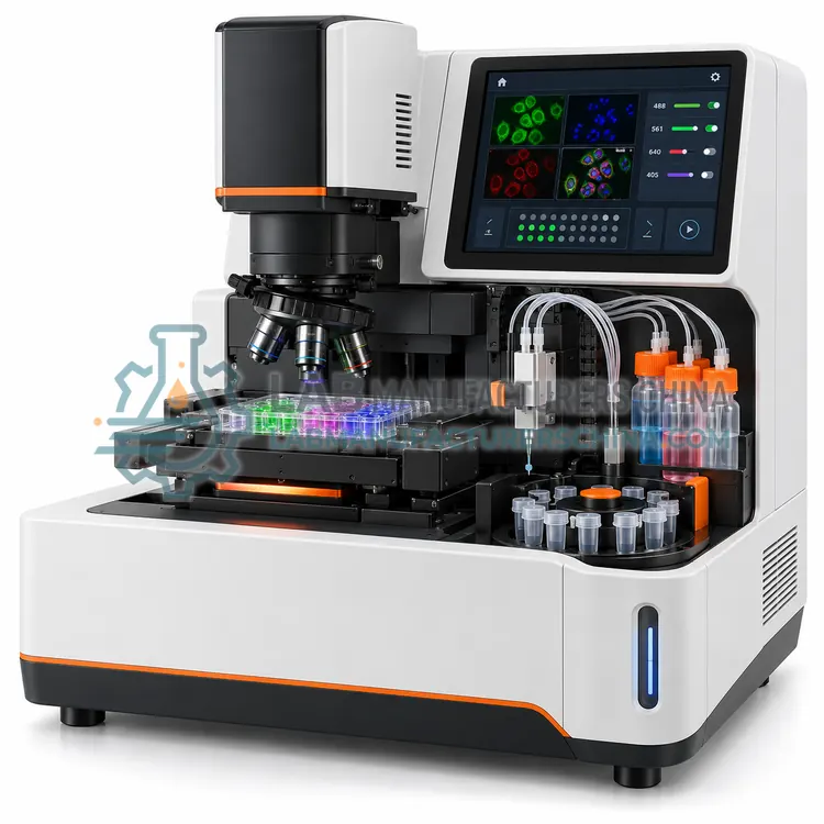
Fluorescence Cell Imager

荧光细胞成像仪

  • CEI-0005
  • Cell Imaging
  • CE (to be confirmed)
  • ISO 9001 manufacturing (to be confirmed)
  • IEC 61010-1 safety (to be confirmed)
Magnification:
Fixed 4x or 10x equivalent
Imaging channels:
Brightfield plus 2–4 fluorescence channels
Excitation:
approx. 365, 470 and 550 nm options
Camera:
Colour and monochrome CMOS, 5–12 MP
Vessel compatibility:
Flasks, dishes, multi-well plates and slides
Confluence measurement:
0–100% from brightfield contrast

A benchtop fluorescence cell imager is a compact instrument with fixed optics and pre-set channels that captures brightfield and fluorescence images of cells in plates, giving quick visual confirmation of confluence, viability and transfection without a full microscope setup.

Request a quote
View details

Compare Cell Imaging models

ModelObjectivesCameraIncubationData outputDimensions (W x D x H)
Inverted Fluorescence Microscope for Cell CultureCEI-00014x, 10x, 20x, 40x and 60x optionssCMOS or CCD, 4–20 MPOptional stage incubator for temperature and CO2 controlTIFF images with acquisition metadataapprox. 300 x 500 x 500 mm
Confocal Laser Scanning MicroscopeCEI-000210x to 100x with numerical aperture up to 1.4———approx. 600 x 900 x 600 mm
Live-Cell Imaging Incubation SystemCEI-0003————approx. 300 x 400 x 150 mm (chamber)
High-Content Screening Imaging SystemCEI-0004——Controlled temperature, CO2 and humidityPlate-level CSV with per-cell feature tablesapprox. 900 x 700 x 700 mm
Fluorescence Cell ImagerCEI-0005—Colour and monochrome CMOS, 5–12 MP—PNG or TIFF images with confluence and count valuesapprox. 200 x 250 x 200 mm

Indicative values for this product class. Final specifications are confirmed on the quotation and in the calibration documents supplied with the instrument.

How do Cell Imaging work?

All cell imaging systems collect light from a specimen and form an image on a detector. In widefield microscopy, the whole field is illuminated and the image is limited by out-of-focus light, so thin cells look sharp while thick specimens appear hazy. Confocal microscopy places a pinhole in front of the detector so that only light from the focal plane reaches it, and it scans point by point or line by line to build an optical section; this improves contrast and allows three-dimensional reconstruction. Fluorescence excitation comes from LEDs, lasers or arc lamps, and dichroic mirrors and emission filters separate the excitation from the emitted signal. Live-cell systems add an incubated stage or chamber to hold temperature, CO2 and humidity, and software captures time-lapse sequences. High-content systems combine automated plate handling, autofocus and image analysis to extract numerical features from thousands of images.

Which Cell Imaging variants are available?

  • Inverted Fluorescence Microscope for Cell Culture — An inverted fluorescence microscope views cells from below through the base of a culture vessel, with LED excitation and two to five filter sets for routine morphology and immunofluorescence.
  • Confocal Laser Scanning Microscope — A confocal laser scanning microscope uses a pinhole and scanning laser beams to reject out-of-focus light, producing optical sections through thick cells and tissue and enabling high-contrast three-dimensional reconstruction of subcellular structures.
  • Live-Cell Imaging Incubation System — A live-cell imaging incubation system maintains temperature, carbon dioxide, humidity and often oxygen on the microscope stage while the software captures time-lapse sequences, so cell behaviour can be recorded continuously for hours or days.
  • High-Content Screening Imaging System — A high-content screening imaging system automates plate handling, autofocus, multi-channel image acquisition and analysis, extracting quantitative single-cell features from thousands of wells for compound screening, dose-response studies and phenotypic assay development in drug discovery.
  • Fluorescence Cell Imager — A benchtop fluorescence cell imager is a compact instrument with fixed optics and pre-set channels that captures brightfield and fluorescence images of cells in plates, giving quick visual confirmation of confluence, viability and transfection without a full microscope setup.

How do I choose the right variant?

Match the system to the spatial and temporal resolution the experiment needs. Routine morphology, transfection checks and immunofluorescence on coverslips are well served by an inverted fluorescence microscope with LED illumination and a scientific camera. Confocal systems are needed when optical sections, thick specimens, three-dimensional reconstruction or low background matter, and spinning-disk confocals add speed for live imaging. Live-cell work requires an incubation chamber with CO2 and temperature control, plus low phototoxicity and a camera sensitive enough to limit exposure. High-content platforms are chosen when hundreds to thousands of wells must be imaged and quantified automatically. Check objective quality and numerical aperture, available fluorescence channels, detector sensitivity, stage automation, autofocus reliability, incubation performance and the analysis software, which often matters more than optics for quantitative results.

Where are Cell Imaging used?

Cell Imaging are specified in cell morphology and confluence assessment, immunofluorescence and protein localisation, live-cell dynamics and time-lapse studies, high-content compound screening and transfection and reporter gene analysis.

Which standards and certifications apply?

Work with Cell Imaging is commonly governed by IEC 60825-1, IEC 61010-1, IEC 61010-2-010, ISO 17025 and ISO 20387. Instrument certifications such as CE marking are confirmed per configuration and never assumed.

What are the key considerations before purchase?

Optical cleanliness is essential, so objectives should be cleaned with lens tissue and the recommended solvent, and immersion oil removed after every session. Live-cell imaging requires careful control of temperature, CO2 and humidity, because evaporation changes osmolarity and focus drift spoils long time-lapse runs; hardware autofocus or a focus-maintenance routine is usually needed. Phototoxicity and photobleaching limit live experiments, so use the lowest illumination power and shortest exposure that gives an acceptable signal, and reduce the number of channels and time points. Laser systems fall under IEC 60825-1 and must remain interlocked. After installation, verify alignment with a calibration slide, then check stage repeatability and focus stability periodically, and keep records so that images from different sessions remain comparable.

Frequently asked questions about Cell Imaging

Confocal is needed when out-of-focus light obscures the signal, for example in thick specimens, three-dimensional reconstructions, thick tissue and sections with high background. For thin cultured cells and bright markers, widefield imaging with a good camera is often sufficient.

Need help specifying Cell Imaging?

Request a quote