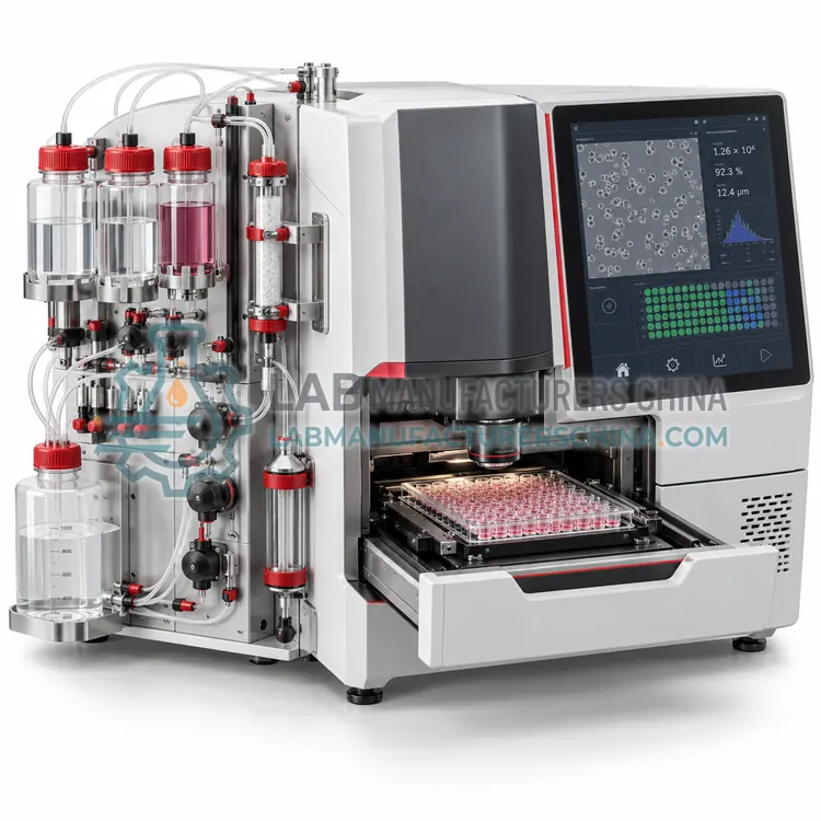
How do Cell Counters work?
Counting methods differ in how they distinguish a cell from debris and from a dead cell. Trypan blue exclusion relies on the intact membrane of viable cells, which excludes the dye, so live cells appear bright and dead cells blue in transmitted light; automated counters image a fixed volume in a counting chamber and classify objects by size, shape and brightness. Fluorescence methods use acridine orange and propidium iodide, which stain all nuclei and membrane-damaged nuclei respectively, so concentration and viability are derived from two channels and are less affected by red blood cells, platelets and debris. Electrical impedance counters draw cells through a small aperture and count voltage pulses, where pulse height reflects cell volume. Flow cytometry counters add a known number of reference beads and calculate absolute counts from the ratio of cell to bead events, which also provides marker information. All methods require a well-mixed, appropriately diluted sample to be accurate.
Which Cell Counters variants are available?
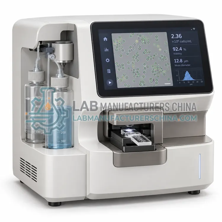
- Automated Image-Based Cell Counter — An automated image-based cell counter loads a small sample into a disposable slide, images a defined volume in transmitted light and reports concentration and viability from trypan blue exclusion.
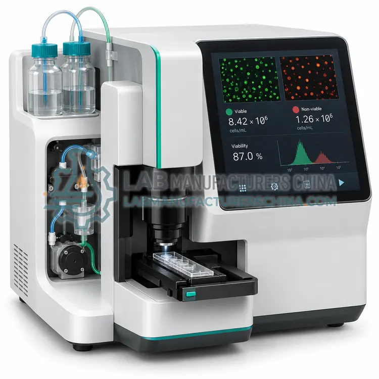
- Dual-Fluorescence Cell Counter — A dual-fluorescence cell counter stains nuclei with acridine orange and propidium iodide and measures both channels, reporting concentration and viability even in samples containing red blood cells, platelets or debris.
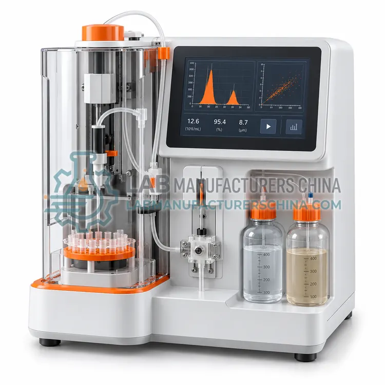
- Electrical Impedance Cell Counter — An electrical impedance cell counter draws cells through a small aperture and counts voltage pulses whose height reflects cell volume, giving concentration and size distribution without any dye or optical measurement.
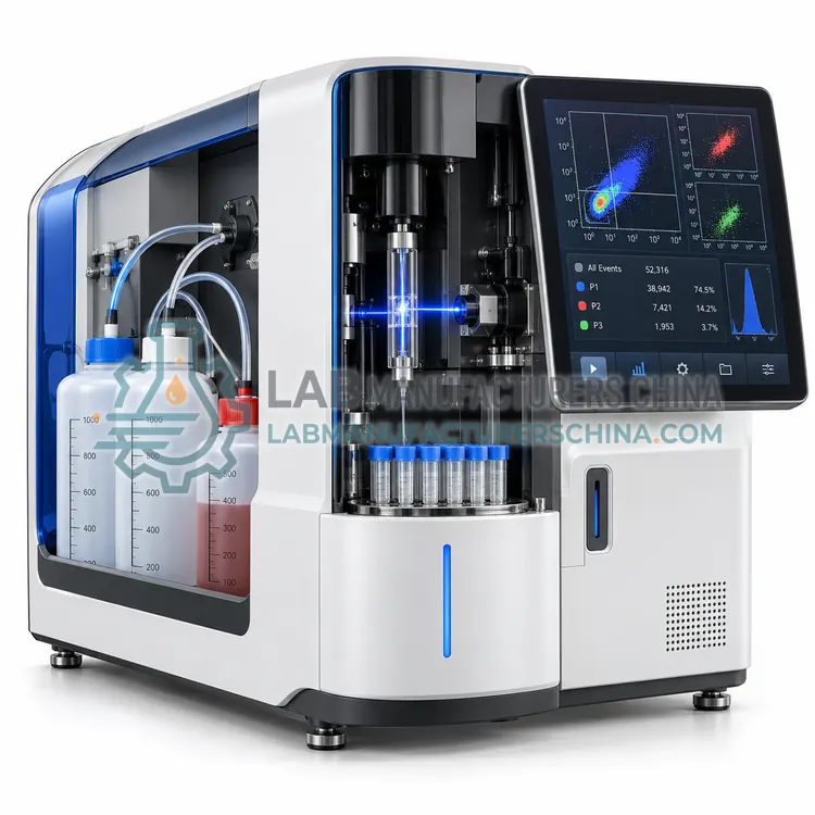
- Flow Cytometry Cell Counter — A flow cytometry cell counter mixes the sample with a known number of reference beads and calculates absolute counts from the ratio of cell to bead events, combining concentration measurement with marker and viability information in one acquisition.
- High-Throughput Automated Cell Counter — A high-throughput automated cell counter reads multi-well plates and reports concentration and viability well by well, processing a 96-well plate in minutes for bioprocess development, clone screening and cell-based assay quality control work.
How do I choose the right variant?
Choose by sample type and required accuracy. For routine culture of established cell lines, an image-based counter with trypan blue is adequate and inexpensive. When samples contain debris, red cells, platelets or clumps, or when viability is low and must be measured precisely, choose a fluorescence counter with acridine orange and propidium iodide, which reports concentration and viability from nuclear staining. Impedance counters are useful for continuous monitoring of cell size distribution and for samples where optical clarity varies. Flow cytometry counters are the right choice when counts must be combined with surface marker measurement or when absolute counts are required for clinical reporting. Check the linear range, sample volume, time per count, consumable cost, cleaning requirements and whether the instrument reports aggregate or clump correction.
Where are Cell Counters used?
Cell Counters are specified in seeding density determination before experiments, culture growth and viability monitoring, cell therapy product release counts, bioreactor inoculation and harvest checks and transfection efficiency normalisation.
Which standards and certifications apply?
Work with Cell Counters is commonly governed by ISO 20387, ISO 15189, IEC 61010-1, IEC 60825-1, USP <1046> and ISO 17025. Instrument certifications such as CE marking are confirmed per configuration and never assumed.
What are the key considerations before purchase?
Accuracy depends more on sample handling than on the instrument. Cells settle quickly, so mix by gentle inversion or low-speed pipetting immediately before sampling, and avoid vigorous vortexing that damages membranes. Dilute into the method's linear range, since concentrated samples saturate the sensor and very dilute samples lose precision. Consumables such as counting slides or chambers should be used once and disposed of as biohazard waste. Clean the optics and sample path regularly, and avoid samples containing strong dyes or fluorescent compounds that interfere with fluorescence channels. Validate each new cell type or matrix against a reference method such as a haemocytometer or a validated flow count, and document the dilution and settings used, because viability definitions differ between laboratories.