High-Throughput Automated Cell Counter
高通量自动细胞计数仪
- SKU CEC-0005
- Cell Counters
- CE (to be confirmed)
- ISO 9001 manufacturing (to be confirmed)
- IEC 61010-1 safety (to be confirmed)
Price on request — FOB / CIF quoted per order
Prices depend on configuration, quantity and destination port.
Key specifications
| Plate formats | 96-well and 24-well |
|---|---|
| Plate processing time | approx. 5–15 min |
| Sample volume per well | 10–20 µL |
| Concentration range | approx. 1 x 10⁴–1 x 10⁷ cells/mL |
| Cell size range | approx. 3–60 µm |
| Imaging modes | Brightfield, optional fluorescence channels |
Request a quote Download brochure · to be provided
By Lab Manufacturers China Technical TeamLast reviewed Standards: ISO 20387, ISO 15189, IEC 61010-1
What is the High-Throughput Automated Cell Counter?
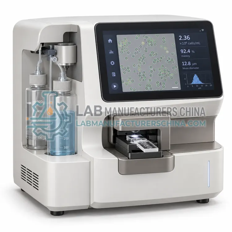
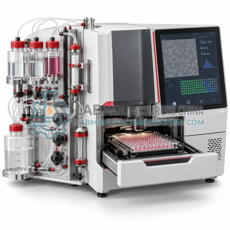
A high-throughput automated cell counter reads multi-well plates and reports concentration and viability well by well, processing a 96-well plate in minutes for bioprocess development, clone screening and cell-based assay quality control work.
What is the High-Throughput Automated Cell Counter?
A high-throughput automated cell counter reads multi-well plates and reports concentration and viability well by well, processing a 96-well plate in minutes for bioprocess development, clone screening and cell-based assay quality control work.
Overview
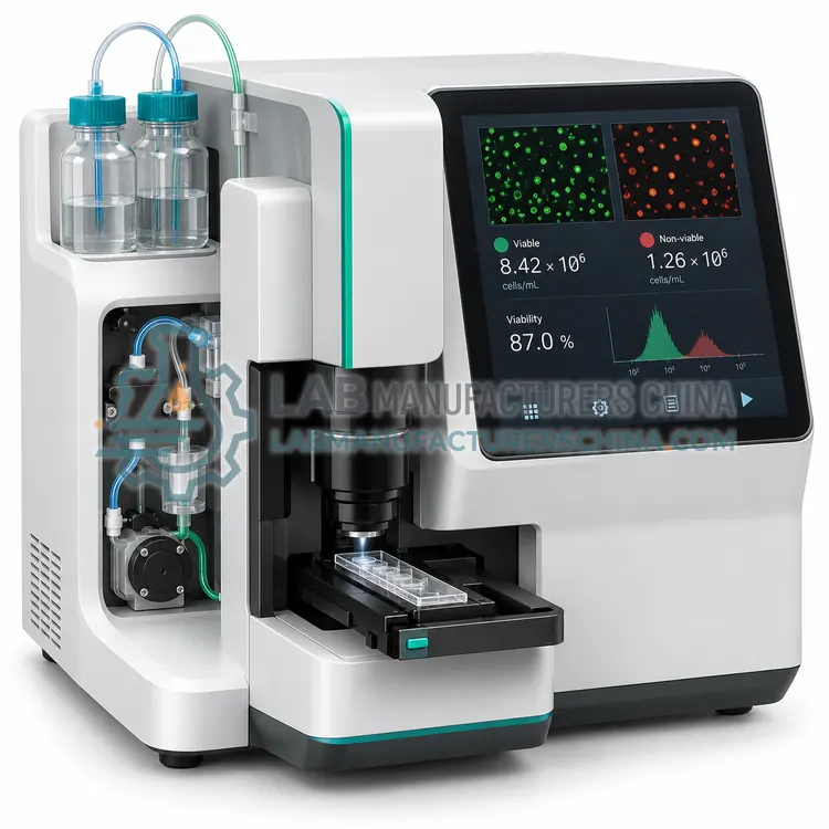
The instrument combines a plate handler, brightfield and fluorescence optics and software that analyses each well independently. Plates are prepared by mixing a small volume of cell suspension with viability dye, and the counter images a defined area in every well, reporting concentration, viability, mean cell diameter and, where fluorescence channels are fitted, cell counts distinguished by fluorescent label. A full 96-well plate is typically processed in 5–15 minutes, so time-course or clone-screening experiments can be counted repeatedly without manual handling. Because sample volumes are small, 10–20 µL per well is sufficient. Cross-contamination between wells is limited by disposable plates and a fixed optical stage, and results are exported as a plate map for further analysis.
How does it work?
Counting methods differ in how they distinguish a cell from debris and from a dead cell. Trypan blue exclusion relies on the intact membrane of viable cells, which excludes the dye, so live cells appear bright and dead cells blue in transmitted light; automated counters image a fixed volume in a counting chamber and classify objects by size, shape and brightness. Fluorescence methods use acridine orange and propidium iodide, which stain all nuclei and membrane-damaged nuclei respectively, so concentration and viability are derived from two channels and are less affected by red blood cells, platelets and debris. Electrical impedance counters draw cells through a small aperture and count voltage pulses, where pulse height reflects cell volume. Flow cytometry counters add a known number of reference beads and calculate absolute counts from the ratio of cell to bead events, which also provides marker information. All methods require a well-mixed, appropriately diluted sample to be accurate.
What are the key features?
- Multi-well plate reading with per-well results
- Brightfield and optional fluorescence channels
- 96-well plate processed in 5–15 minutes
- Reported concentration, viability and mean diameter
- Plate map export for time-course or clone screening
- Low sample consumption of 10–20 µL per well
What are the technical specifications?
A typical high-throughput automated cell counter configuration offers plate formats of 96-well and 24-well, plate processing time of approx. 5–15 min, sample volume per well of 10–20 µL, concentration range of approx. 1 x 10⁴–1 x 10⁷ cells/mL, cell size range of approx. 3–60 µm and imaging modes of Brightfield, optional fluorescence channels. Values are indicative of the product class; the exact figures for each configuration are confirmed on the quotation and the calibration documents supplied with the instrument.
Which applications is it used for?
The High-Throughput Automated Cell Counter is used in clone screening and clone stability studies, bioreactor time-course cell density monitoring, cell-based assay plate quality control and media and supplement optimisation experiments. Across the wider Cell Counters class, laboratories also deploy it for seeding density determination before experiments, culture growth and viability monitoring and cell therapy product release counts.
Which standards and certifications apply?
Relevant standards and methods include ISO 20387, ISO 15189, IEC 61010-1, IEC 60825-1, USP <1046> and ISO 17025. Certification candidates for this model are CE, ISO 9001 manufacturing and IEC 61010-1 safety; each is to be confirmed for the specific configuration before shipment, and certificates are supplied only where they have been issued.
Accuracy depends more on sample handling than on the instrument. Cells settle quickly, so mix by gentle inversion or low-speed pipetting immediately before sampling, and avoid vigorous vortexing that damages membranes. Dilute into the method's linear range, since concentrated samples saturate the sensor and very dilute samples lose precision. Consumables such as counting slides or chambers should be used once and disposed of as biohazard waste. Clean the optics and sample path regularly, and avoid samples containing strong dyes or fluorescent compounds that interfere with fluorescence channels. Validate each new cell type or matrix against a reference method such as a haemocytometer or a validated flow count, and document the dilution and settings used, because viability definitions differ between laboratories.
How do I select the right High-Throughput Automated Cell Counter?
Select a plate-based counter when many parallel cultures must be measured repeatedly, such as clone selection or media optimisation. Specify plate formats, processing time, fluorescence channel options, sample volume and whether per-well images are archived for review.
How does it compare with other Cell Counters variants?
Choose by sample type and required accuracy. For routine culture of established cell lines, an image-based counter with trypan blue is adequate and inexpensive. When samples contain debris, red cells, platelets or clumps, or when viability is low and must be measured precisely, choose a fluorescence counter with acridine orange and propidium iodide, which reports concentration and viability from nuclear staining. Impedance counters are useful for continuous monitoring of cell size distribution and for samples where optical clarity varies. Flow cytometry counters are the right choice when counts must be combined with surface marker measurement or when absolute counts are required for clinical reporting. Check the linear range, sample volume, time per count, consumable cost, cleaning requirements and whether the instrument reports aggregate or clump correction.
Full specifications
| Plate formats | 96-well and 24-well |
|---|---|
| Plate processing time | approx. 5–15 min |
| Sample volume per well | 10–20 µL |
| Concentration range | approx. 1 x 10⁴–1 x 10⁷ cells/mL |
| Cell size range | approx. 3–60 µm |
| Imaging modes | Brightfield, optional fluorescence channels |
| Viability dyes | Trypan blue, acridine orange and propidium iodide |
| Data output | Plate map CSV with per-well values |
| Ambient control | Not required; closed imaging chamber |
| Dimensions (W x D x H) | approx. 500 x 550 x 400 mm |
Indicative values for this product class. Final specifications are confirmed on the quotation and in the calibration documents supplied with the instrument.
Applications
- Clone screening and clone stability studies
- Bioreactor time-course cell density monitoring
- Cell-based assay plate quality control
- Media and supplement optimisation experiments
Key features
- Multi-well plate reading with per-well results
- Brightfield and optional fluorescence channels
- 96-well plate processed in 5–15 minutes
- Reported concentration, viability and mean diameter
- Plate map export for time-course or clone screening
- Low sample consumption of 10–20 µL per well
Frequently asked questions about High-Throughput Automated Cell Counter
Typically 5–15 minutes depending on the number of imaging channels and the field of view per well. That makes repeated counting of a time-course plate practical without interrupting the cultures for long.
Yes on models with fluorescence channels. Labels such as GFP or fluorescent antibodies allow labelled cells to be counted separately from unlabelled ones, so transfection efficiency or population ratios can be monitored across the plate.
Most measurements are short enough to be performed at room temperature without a controlled chamber, but plates should be returned to the incubator promptly and repeated counts should follow the same timing to avoid temperature-related variation.
Most benchtop counters use 10–20 µL per count, and some fluorescence systems accept as little as 10 µL when viability is measured from nuclear staining. Samples are usually diluted with dye or buffer before counting.
Trypan blue measures membrane integrity, whereas fluorescence nuclear dyes also detect early apoptosis and can include cells that have lost nuclear structure. Instruments also differ in how they classify small or clumped objects, so methods should not be mixed within a study.
Gently pipette the suspension with a wide-bore tip or filter through a 40 µm mesh to break clumps before counting. Some instruments apply a clump correction factor, which should be verified against a reference count for each cell type.
Related models