How do Protein Analyzers work?
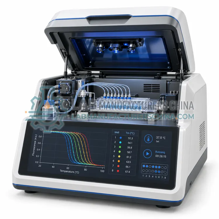
Protein analyzers apply a physical or chemical measurement that responds to a specific property of the molecule. Amino acid analysis hydrolyses the protein and separates the released amino acids by ion exchange or reversed-phase chromatography with ninhydrin or fluorescence detection, giving composition in residues per molecule. Edman degradation removes one amino acid at a time from the N-terminus by phenylisothiocyanate chemistry and identifies each derivative chromatographically, giving the first 20–50 residues. Circular dichroism measures the difference in absorption of left- and right-circularly polarised light, and the far-UV spectrum reports alpha helix, beta sheet and random coil content. Thermal shift assays use a fluorescent dye that reports unfolding as temperature rises, giving a melting temperature that shifts when a ligand binds. Surface plasmon resonance detects mass binding at a sensor surface in real time, yielding association and dissociation rate constants.
Which Protein Analyzers variants are available?
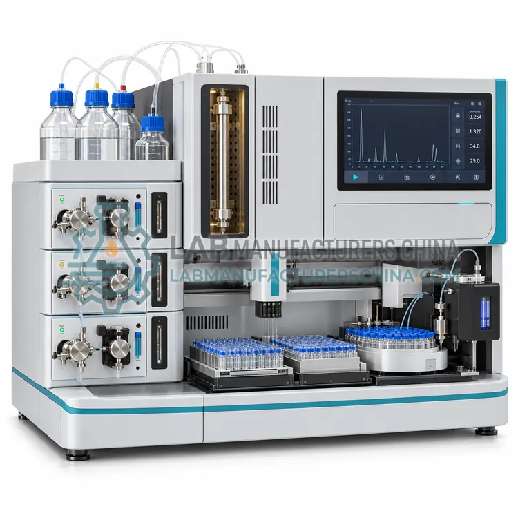
- Automated Amino Acid Analyzer — An automated amino acid analyzer hydrolyses a protein sample and quantifies the released amino acids by ion exchange or reversed-phase separation with post-column or pre-column derivatisation.
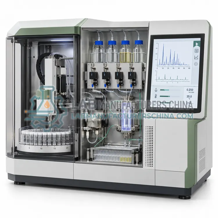
- Protein Sequencer — A protein sequencer determines the N-terminal amino acid sequence of a purified peptide or protein by automated Edman degradation, identifying 20 to 50 residues per run.
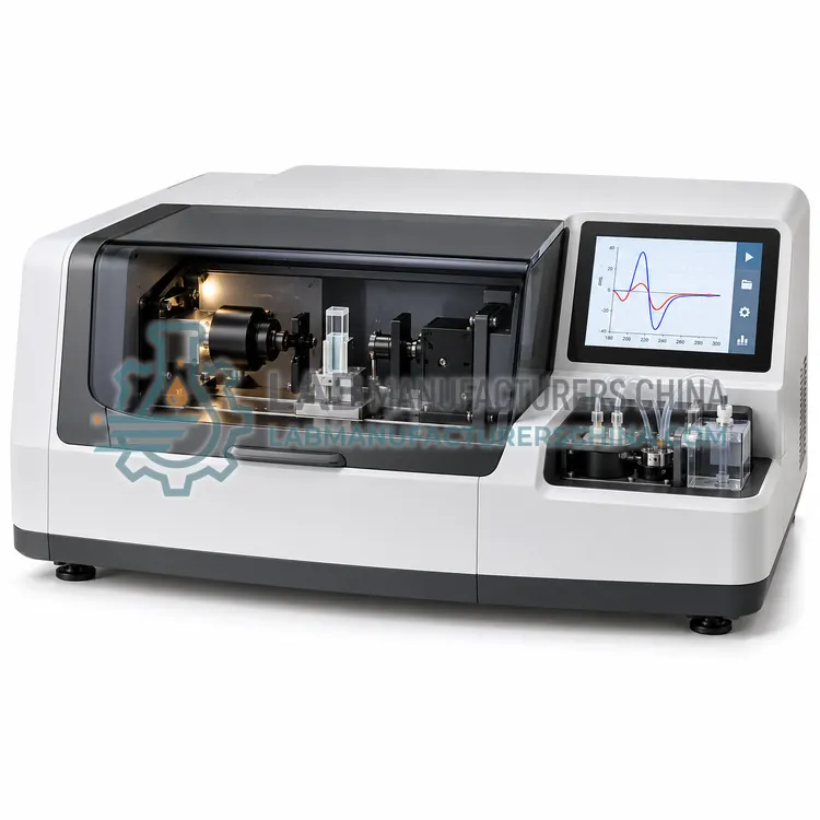
- Circular Dichroism Spectrometer — A circular dichroism spectrometer measures the difference in absorption of left- and right-circularly polarised light to report protein secondary structure, folding state and thermal unfolding.
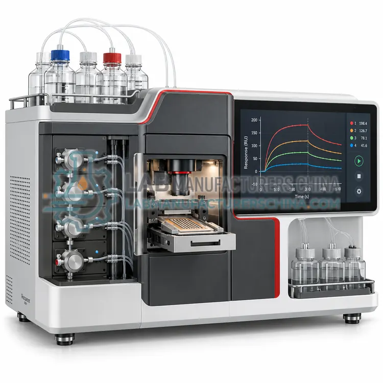
- Surface Plasmon Resonance Analyzer — A surface plasmon resonance analyzer measures binding between molecules in real time by detecting mass changes at a sensor surface, giving association and dissociation rate constants and affinity.
- Protein Thermal Shift Analyzer — A protein thermal shift analyzer measures the melting temperature of a protein in solution using a fluorescent dye that reports unfolding, screening buffer conditions and ligand binding in 96-well format.
How do I choose the right variant?
Choose the analyzer that measures the property you must document. For composition or identity of a purified protein, amino acid analysis gives a quantitative profile; Edman sequencing provides N-terminal sequence but needs a pure, unblocked sample. Circular dichroism is the standard method for secondary structure comparison between batches and for folding studies, and it needs only 0.2–0.5 mg/mL in a low-absorbing buffer. Thermal shift assays screen buffer conditions and ligand binding quickly in 96-well format, making them useful early in development. Surface plasmon resonance quantifies affinity and kinetics, and it demands careful immobilisation chemistry and reference channel design. Compare sample consumption, sensitivity, throughput, buffer tolerance, temperature control and whether the software can export raw traces for independent analysis.
Where are Protein Analyzers used?
Protein Analyzers are specified in recombinant protein characterisation, biosimilar comparability studies, protein structure and folding research, ligand binding and affinity measurement and protein identity confirmation.
Which standards and certifications apply?
Work with Protein Analyzers is commonly governed by USP <1057>, Ph. Eur. 2.2.56, Ph. Eur. 2.2.25, ISO 17025, IEC 61010-1 and 21 CFR Part 11. Instrument certifications such as CE marking are confirmed per configuration and never assumed.
What are the key considerations before purchase?
Sample purity and buffer composition dominate results, so prepare proteins in volatile, low-absorbing buffers for circular dichroism and avoid detergents and reducing agents that absorb in the far UV. Amino acid analysis needs complete hydrolysis, so ampoule sealing, vacuum and hydrolysis time must be controlled. Surface plasmon resonance sensors are sensitive to buffer refractive index differences, so running buffer and sample buffer should match and a reference channel should be included. Thermal shift assays need consistent dye concentration and controlled heating rates. Verify performance periodically with a reference protein such as bovine serum albumin or lysozyme, keep maintenance and calibration records, and store raw data, since circular dichroism and biosensor results are often re-analysed after method changes.