How do Protein Electrophoresis work?
In SDS-PAGE, the sample is denatured with sodium dodecyl sulfate and a reducing agent so that proteins unfold and acquire a uniform negative charge proportional to their length. When an electric field is applied, all proteins migrate toward the anode, and the polyacrylamide mesh sieves them so that migration distance depends mainly on molecular weight. A discontinuous buffer system with stacking and resolving gel layers concentrates proteins into a sharp band before separation. Isoelectric focusing instead uses a pH gradient and separates proteins according to their isoelectric point, where net charge is zero and migration stops. Two-dimensional electrophoresis combines the two, separating by charge first and by size second, which resolves thousands of protein spots. Capillary systems perform SDS or charge separation in a narrow capillary with on-line absorbance or fluorescence detection, replacing staining with quantitative detection.
Which Protein Electrophoresis variants are available?
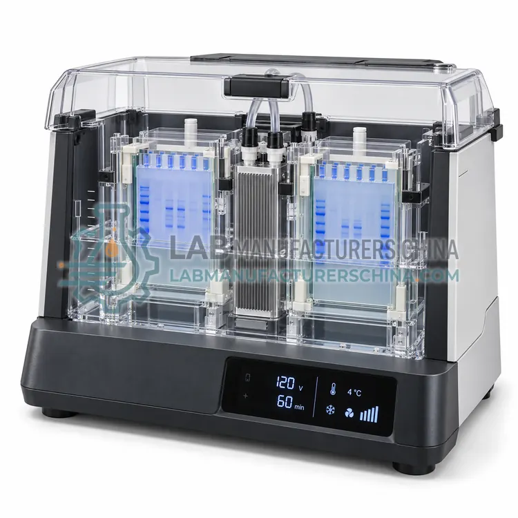
- Vertical SDS-PAGE Electrophoresis System — A vertical SDS-PAGE system runs one or two 1 mm polyacrylamide gels in a buffer tank with platinum wire electrodes, resolving proteins from 10 to 200 kDa.
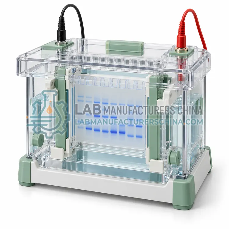
- Mini Vertical Gel Electrophoresis System — A mini vertical electrophoresis system uses smaller 8 x 8 cm gels and roughly 200 mL of buffer, completing a separation in 30 to 60 minutes with low sample and reagent consumption.
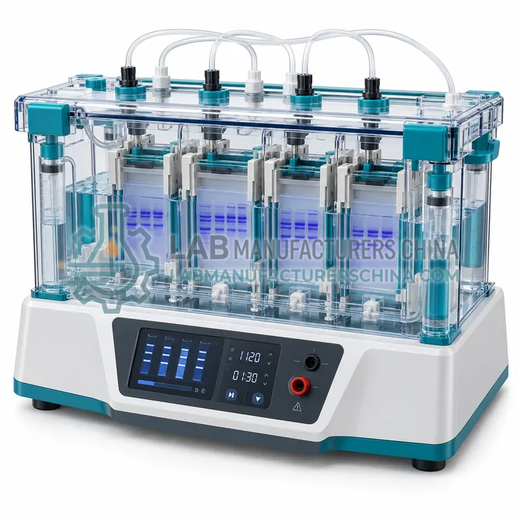
- Multichannel Vertical Electrophoresis System — A multichannel vertical system runs 6 to 12 gels in one tank with shared buffer chambers and uniform electrode geometry, screening large sample sets under identical conditions in a single run.
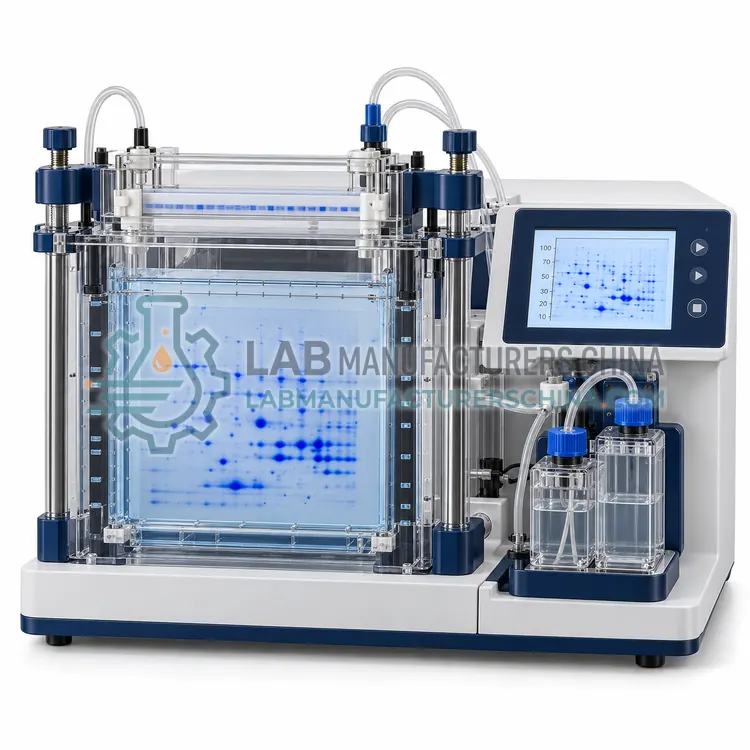
- Two-Dimensional Electrophoresis System — A two-dimensional electrophoresis system separates proteins first by isoelectric point in an immobilised pH gradient strip and then by molecular weight in a polyacrylamide gel, resolving thousands of protein spots from complex lysates for comparative proteome analysis.
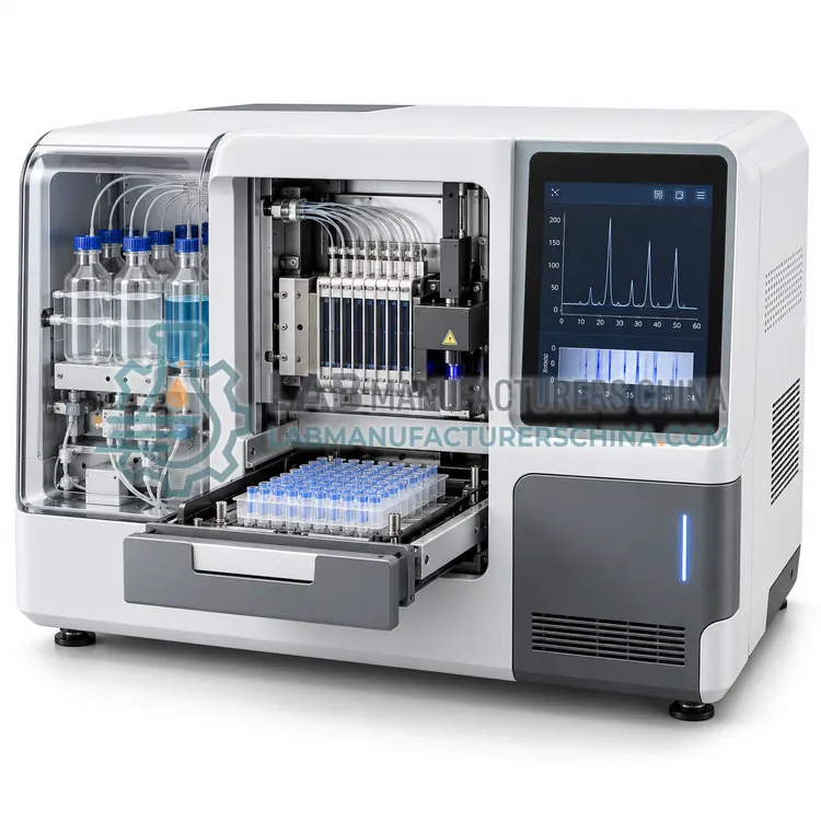
- Automated Capillary Protein Electrophoresis System — An automated capillary protein electrophoresis system performs SDS or charge-based separation in capillaries with on-line absorbance detection, reporting purity as a numerical percentage.
How do I choose the right variant?
Start with the purpose. For routine purity and molecular weight, a vertical SDS-PAGE system with 1 mm gels and 10–15 wells covers most needs and is inexpensive. Mini systems reduce buffer and sample consumption, run faster and are convenient for immunoblotting, at the cost of lower resolution for closely spaced bands. Multichannel systems process 6 to 12 gels in one tank when screening many samples. Two-dimensional electrophoresis is chosen when complex proteomes must be resolved, and it requires an isoelectric focusing first dimension plus consistent sample preparation. Capillary systems give numerical purity results and are preferred where regulated release testing, low sample volume or full automation is required. Check gel percentage and gradient availability, maximum voltage and current, cooling provisions, glass plate compatibility and whether precast gels are supported.
Where are Protein Electrophoresis used?
Protein Electrophoresis are specified in protein purity and molecular weight determination, recombinant protein expression analysis, two-dimensional proteome profiling, monoclonal protein detection in clinical samples and blot preparation for immunodetection.
Which standards and certifications apply?
Work with Protein Electrophoresis is commonly governed by USP <1057>, Ph. Eur. 2.2.31, Ph. Eur. 2.2.34, IEC 61010-1, ISO 17025 and ISO 15189. Instrument certifications such as CE marking are confirmed per configuration and never assumed.
What are the key considerations before purchase?
Acrylamide is a neurotoxin and suspected carcinogen, so powder handling and gel casting should be performed in a fume hood or with ready-to-use precast gels, and gloves must be worn. Buffer pH and ionic strength affect stacking and resolution, so working buffers should be prepared fresh and not reused excessively. Consistent sample loading, reducing agent freshness and heating time determine how sharp bands appear. Gels should be run within the recommended voltage and current limits, and temperature control matters for high-voltage runs because Joule heating distorts bands. Staining and destaining protocols must be standardised for comparative densitometry, and images used for purity calculations should be captured in the linear range of the camera. Keep records of gel percentage, running conditions and staining for traceability.