Automated Capillary Protein Electrophoresis System
自动化毛细管蛋白质电泳系统
- SKU PRE-0005
- Protein Electrophoresis
- CE (to be confirmed)
- ISO 9001 manufacturing (to be confirmed)
- IEC 61010-1 safety (to be confirmed)
- 21 CFR Part 11 software compliance (to be confirmed)
Price on request — FOB / CIF quoted per order
Prices depend on configuration, quantity and destination port.
Key specifications
| Separation modes | SDS gel or free-solution charge based |
|---|---|
| Detection | UV absorbance at 200 or 214 nm |
| Molecular weight range | approx. 10–225 kDa |
| Purity resolution | approximately 0.1% peak area |
| Run time per sample | approx. 10–40 min |
| Sample volume | 20–100 µL of diluted sample |
Request a quote Download brochure · to be provided
By Lab Manufacturers China Technical TeamLast reviewed Standards: USP <1057>, Ph. Eur. 2.2.31, Ph. Eur. 2.2.34
What is the Automated Capillary Protein Electrophoresis System?
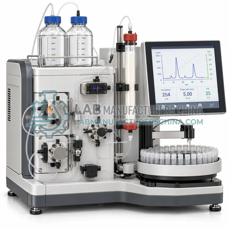
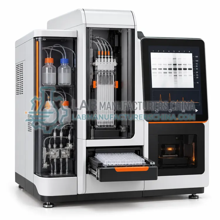
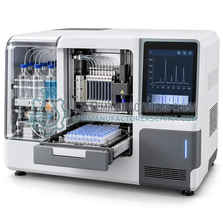
An automated capillary protein electrophoresis system performs SDS or charge-based separation in capillaries with on-line absorbance detection, reporting purity as a numerical percentage. It is used for release testing and comparability studies where quantitative, auditable results are required.
What is the Automated Capillary Protein Electrophoresis System?
An automated capillary protein electrophoresis system performs SDS or charge-based separation in capillaries with on-line absorbance detection, reporting purity as a numerical percentage. It is used for release testing and comparability studies where quantitative, auditable results are required.
Overview
The instrument injects a diluted protein sample into a polymer-filled capillary and separates proteins by size in an SDS gel buffer or by charge in a free-solution buffer. On-line UV detection at 200 or 214 nm records the migration of each species, and the software integrates the peaks to give corrected area percentages for each purity group, including fragments and aggregates. Typical sample consumption is 20–100 µL of diluted solution, and a run completes in 10–40 minutes per sample with several capillaries operating in parallel. Reducing and non-reducing conditions can be run in sequence, and molecular weight can be assigned against a protein standard included in every run. Because detection is UV-based and data are digital, results are traceable and suitable for regulated environments.
How does it work?
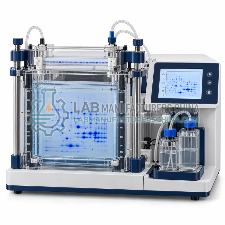
In SDS-PAGE, the sample is denatured with sodium dodecyl sulfate and a reducing agent so that proteins unfold and acquire a uniform negative charge proportional to their length. When an electric field is applied, all proteins migrate toward the anode, and the polyacrylamide mesh sieves them so that migration distance depends mainly on molecular weight. A discontinuous buffer system with stacking and resolving gel layers concentrates proteins into a sharp band before separation. Isoelectric focusing instead uses a pH gradient and separates proteins according to their isoelectric point, where net charge is zero and migration stops. Two-dimensional electrophoresis combines the two, separating by charge first and by size second, which resolves thousands of protein spots. Capillary systems perform SDS or charge separation in a narrow capillary with on-line absorbance or fluorescence detection, replacing staining with quantitative detection.
What are the key features?
- SDS size-based or charge-based separation modes
- On-line UV detection at 200 or 214 nm
- Corrected peak area purity percentages for each species
- Molecular weight assignment against an included standard
- Reduced and non-reduced conditions in one sequence
- Audit trail, user access control and validated software
What are the technical specifications?
A typical automated capillary protein electrophoresis system configuration offers separation modes of SDS gel or free-solution charge based, detection of UV absorbance at 200 or 214 nm, molecular weight range of approx. 10–225 kDa, purity resolution of approximately 0.1% peak area, run time per sample of approx. 10–40 min and sample volume of 20–100 µL of diluted sample. Values are indicative of the product class; the exact figures for each configuration are confirmed on the quotation and the calibration documents supplied with the instrument.
Which applications is it used for?
The Automated Capillary Protein Electrophoresis System is used in biopharmaceutical purity and release testing, biosimilar comparability studies, fragment and aggregate quantification and charge variant profiling of proteins. Across the wider Protein Electrophoresis class, laboratories also deploy it for protein purity and molecular weight determination, recombinant protein expression analysis and two-dimensional proteome profiling.
Which standards and certifications apply?
Relevant standards and methods include USP <1057>, Ph. Eur. 2.2.31, Ph. Eur. 2.2.34, IEC 61010-1, ISO 17025 and ISO 15189. Certification candidates for this model are CE, ISO 9001 manufacturing, IEC 61010-1 safety and 21 CFR Part 11 software compliance; each is to be confirmed for the specific configuration before shipment, and certificates are supplied only where they have been issued.
Acrylamide is a neurotoxin and suspected carcinogen, so powder handling and gel casting should be performed in a fume hood or with ready-to-use precast gels, and gloves must be worn. Buffer pH and ionic strength affect stacking and resolution, so working buffers should be prepared fresh and not reused excessively. Consistent sample loading, reducing agent freshness and heating time determine how sharp bands appear. Gels should be run within the recommended voltage and current limits, and temperature control matters for high-voltage runs because Joule heating distorts bands. Staining and destaining protocols must be standardised for comparative densitometry, and images used for purity calculations should be captured in the linear range of the camera. Keep records of gel percentage, running conditions and staining for traceability.
How do I select the right Automated Capillary Protein Electrophoresis System?
Select a capillary protein electrophoresis system when purity must be reported as a numerical percentage with full traceability, or when sample volume is limited. Specify separation modes, detection wavelength, molecular weight range, capillary count, analysis software and validation documentation.
How does it compare with other Protein Electrophoresis variants?
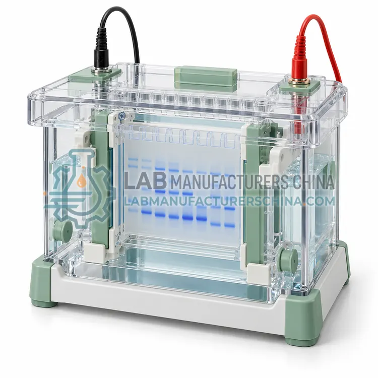
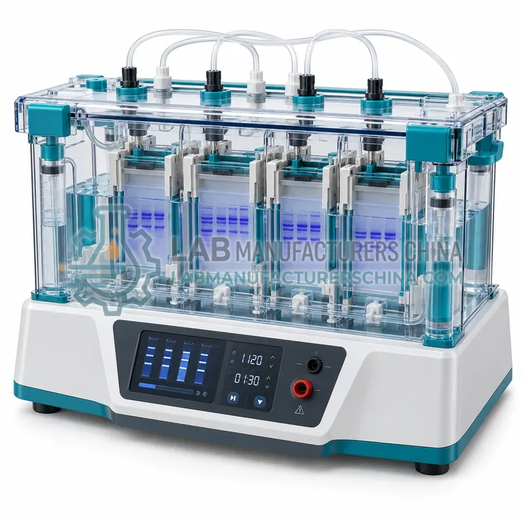
Start with the purpose. For routine purity and molecular weight, a vertical SDS-PAGE system with 1 mm gels and 10–15 wells covers most needs and is inexpensive. Mini systems reduce buffer and sample consumption, run faster and are convenient for immunoblotting, at the cost of lower resolution for closely spaced bands. Multichannel systems process 6 to 12 gels in one tank when screening many samples. Two-dimensional electrophoresis is chosen when complex proteomes must be resolved, and it requires an isoelectric focusing first dimension plus consistent sample preparation. Capillary systems give numerical purity results and are preferred where regulated release testing, low sample volume or full automation is required. Check gel percentage and gradient availability, maximum voltage and current, cooling provisions, glass plate compatibility and whether precast gels are supported.
Full specifications
| Separation modes | SDS gel or free-solution charge based |
|---|---|
| Detection | UV absorbance at 200 or 214 nm |
| Molecular weight range | approx. 10–225 kDa |
| Purity resolution | approximately 0.1% peak area |
| Run time per sample | approx. 10–40 min |
| Sample volume | 20–100 µL of diluted sample |
| Protein concentration | approx. 0.1–2 mg/mL after dilution |
| Capillaries | 1–8 parallel channels (model dependent) |
| Data output | Electropherograms with corrected area report |
| Dimensions (W x D x H) | approx. 500 x 600 x 500 mm |
Indicative values for this product class. Final specifications are confirmed on the quotation and in the calibration documents supplied with the instrument.
Applications
- Biopharmaceutical purity and release testing
- Biosimilar comparability studies
- Fragment and aggregate quantification
- Charge variant profiling of proteins
Key features
- SDS size-based or charge-based separation modes
- On-line UV detection at 200 or 214 nm
- Corrected peak area purity percentages for each species
- Molecular weight assignment against an included standard
- Reduced and non-reduced conditions in one sequence
- Audit trail, user access control and validated software
Frequently asked questions about Automated Capillary Protein Electrophoresis System
Capillary systems typically resolve peaks differing by about 0.1% of the total area, which is finer than gel densitometry, where 1–5% is a realistic limit. The improved resolution comes from on-line detection without staining or destaining steps.
Yes. Reduced and non-reduced samples can be sequenced in the same batch, with the reduced run reporting subunit purity and the non-reduced run showing intact protein, fragments and aggregates. Both use the same calibrant standard.
Demonstrate accuracy, repeatability, intermediate precision, linearity, limit of quantitation and specificity for each product, and confirm that the software audit trail is enabled and access-controlled. Instrument qualification and periodic performance checks with a reference protein are also required.
SDS binds proteins in proportion to their length and gives them a uniform negative charge, so migration in the gel depends mainly on molecular weight. A stacking gel concentrates the sample into a narrow band before the resolving gel separates the proteins.
Use 15% or gradient gels for proteins below 30 kDa, 12% for 15–80 kDa, 10% for 30–120 kDa and 7.5–8% for proteins above 100 kDa. Gradient gels cover a wide range in one run and are convenient when samples vary.
When a complex mixture such as a cell lysate must be resolved into individual proteins. The first dimension separates by isoelectric point, the second by molecular weight, giving thousands of spots that can be compared between conditions to find differentially expressed proteins.
Related models