Protein Thermal Shift Analyzer
蛋白质热稳定性分析仪
- SKU PAN-0005
- Protein Analyzers
- CE (to be confirmed)
- ISO 9001 manufacturing (to be confirmed)
- IEC 61010-1 safety (to be confirmed)
Price on request — FOB / CIF quoted per order
Prices depend on configuration, quantity and destination port.
Key specifications
| Plate format | 96-well or 384-well |
|---|---|
| Temperature range | approx. 20–95 °C |
| Heating rate | 0.1–5 °C/min (programmable) |
| Temperature accuracy | within ±0.3 °C (typical) |
| Protein consumption | 2–20 µg per well |
| Excitation and emission | approx. 470–490 nm excitation, 610–620 nm emission |
Request a quote Download brochure · to be provided
By Lab Manufacturers China Technical TeamLast reviewed Standards: USP <1057>, Ph. Eur. 2.2.56, Ph. Eur. 2.2.25
What is the Protein Thermal Shift Analyzer?
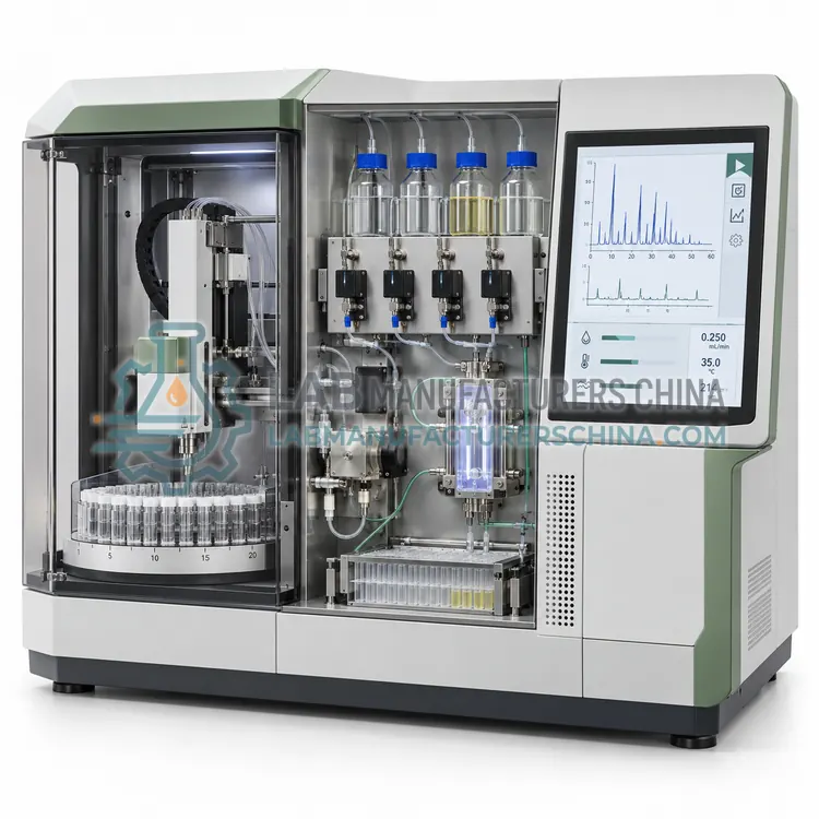
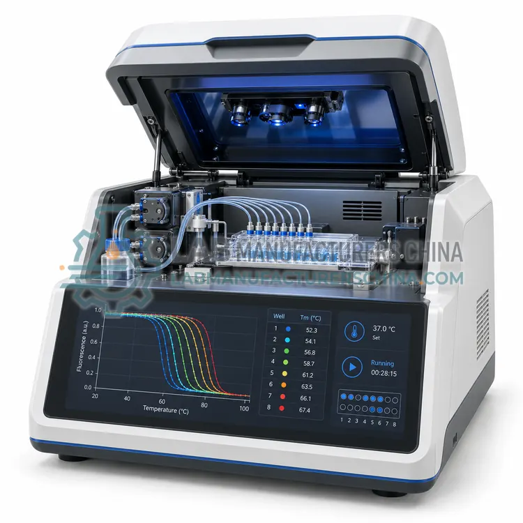
A protein thermal shift analyzer measures the melting temperature of a protein in solution using a fluorescent dye that reports unfolding, screening buffer conditions and ligand binding in 96-well format. It provides fast comparative stability data before detailed structural work.
What is the Protein Thermal Shift Analyzer?
A protein thermal shift analyzer measures the melting temperature of a protein in solution using a fluorescent dye that reports unfolding, screening buffer conditions and ligand binding in 96-well format. It provides fast comparative stability data before detailed structural work.
Overview
The instrument heats a plate containing protein, buffer and a dye whose fluorescence increases when it binds exposed hydrophobic surfaces during unfolding. Fluorescence is recorded as temperature rises from about 20 to 95 °C, and the melting temperature is taken from the inflection point of the resulting curve. Because the readout is comparative, the method ranks conditions rather than giving absolute thermodynamic values, and a stabilising ligand or buffer component appears as an upward shift in melting temperature. Typical protein consumption is 2–20 µg per well, and a full 96-well plate runs in about one to two hours with minimal hands-on work. The same instrument often supports differential scanning fluorimetry for membrane proteins and can be combined with light scattering to detect aggregation during heating.
How does it work?
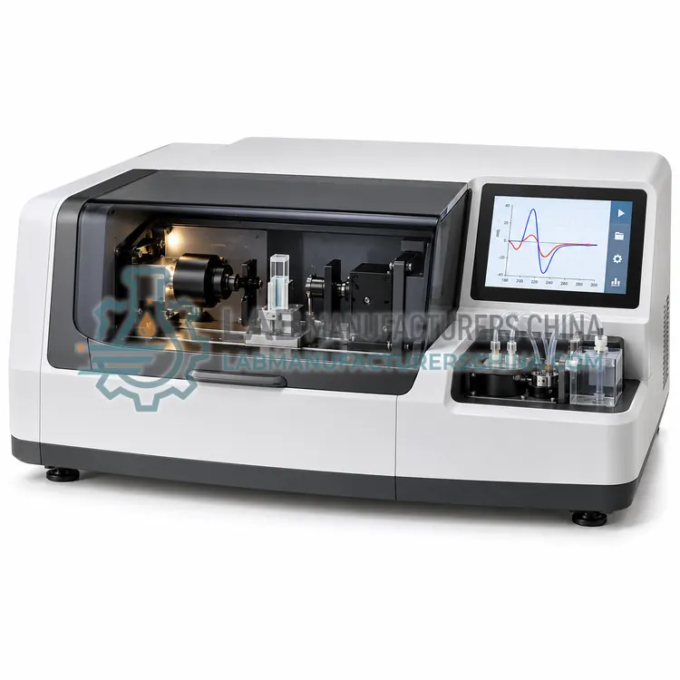
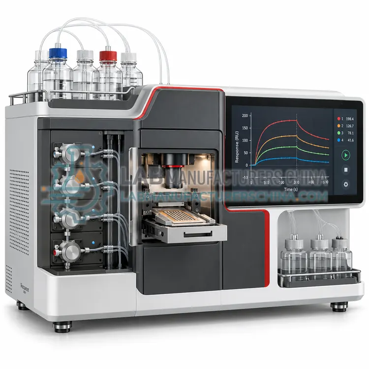
Protein analyzers apply a physical or chemical measurement that responds to a specific property of the molecule. Amino acid analysis hydrolyses the protein and separates the released amino acids by ion exchange or reversed-phase chromatography with ninhydrin or fluorescence detection, giving composition in residues per molecule. Edman degradation removes one amino acid at a time from the N-terminus by phenylisothiocyanate chemistry and identifies each derivative chromatographically, giving the first 20–50 residues. Circular dichroism measures the difference in absorption of left- and right-circularly polarised light, and the far-UV spectrum reports alpha helix, beta sheet and random coil content. Thermal shift assays use a fluorescent dye that reports unfolding as temperature rises, giving a melting temperature that shifts when a ligand binds. Surface plasmon resonance detects mass binding at a sensor surface in real time, yielding association and dissociation rate constants.
What are the key features?
- 96-well plate format with controlled heating to 95 °C
- Melting temperature determination with automatic curve fitting
- Upward shift detection for ligand binding and buffer stabilisation
- Protein consumption of only 2–20 µg per well
- Optional light scattering channel for aggregation monitoring
- Multi-filter detection supporting several dyes
What are the technical specifications?
A typical protein thermal shift analyzer configuration offers plate format of 96-well or 384-well, temperature range of approx. 20–95 °C, heating rate of 0.1–5 °C/min (programmable), temperature accuracy of within ±0.3 °C (typical), protein consumption of 2–20 µg per well and excitation and emission of approx. 470–490 nm excitation, 610–620 nm emission. Values are indicative of the product class; the exact figures for each configuration are confirmed on the quotation and the calibration documents supplied with the instrument.
Which applications is it used for?
The Protein Thermal Shift Analyzer is used in buffer and formulation stability screening, ligand binding confirmation before structural work, protein construct and mutant stability ranking and membrane protein stabilisation studies. Across the wider Protein Analyzers class, laboratories also deploy it for recombinant protein characterisation, biosimilar comparability studies and protein structure and folding research.
Which standards and certifications apply?
Relevant standards and methods include USP <1057>, Ph. Eur. 2.2.56, Ph. Eur. 2.2.25, ISO 17025, IEC 61010-1 and 21 CFR Part 11. Certification candidates for this model are CE, ISO 9001 manufacturing and IEC 61010-1 safety; each is to be confirmed for the specific configuration before shipment, and certificates are supplied only where they have been issued.
Sample purity and buffer composition dominate results, so prepare proteins in volatile, low-absorbing buffers for circular dichroism and avoid detergents and reducing agents that absorb in the far UV. Amino acid analysis needs complete hydrolysis, so ampoule sealing, vacuum and hydrolysis time must be controlled. Surface plasmon resonance sensors are sensitive to buffer refractive index differences, so running buffer and sample buffer should match and a reference channel should be included. Thermal shift assays need consistent dye concentration and controlled heating rates. Verify performance periodically with a reference protein such as bovine serum albumin or lysozyme, keep maintenance and calibration records, and store raw data, since circular dichroism and biosensor results are often re-analysed after method changes.
How do I select the right Protein Thermal Shift Analyzer?
Select a thermal shift analyzer for rapid comparative stability screening when you need to rank buffers, constructs or candidate ligands. Specify plate format, temperature range and heating rate, protein consumption, dye compatibility and whether a light scattering channel is available.
How does it compare with other Protein Analyzers variants?
Choose the analyzer that measures the property you must document. For composition or identity of a purified protein, amino acid analysis gives a quantitative profile; Edman sequencing provides N-terminal sequence but needs a pure, unblocked sample. Circular dichroism is the standard method for secondary structure comparison between batches and for folding studies, and it needs only 0.2–0.5 mg/mL in a low-absorbing buffer. Thermal shift assays screen buffer conditions and ligand binding quickly in 96-well format, making them useful early in development. Surface plasmon resonance quantifies affinity and kinetics, and it demands careful immobilisation chemistry and reference channel design. Compare sample consumption, sensitivity, throughput, buffer tolerance, temperature control and whether the software can export raw traces for independent analysis.
Full specifications
| Plate format | 96-well or 384-well |
|---|---|
| Temperature range | approx. 20–95 °C |
| Heating rate | 0.1–5 °C/min (programmable) |
| Temperature accuracy | within ±0.3 °C (typical) |
| Protein consumption | 2–20 µg per well |
| Excitation and emission | approx. 470–490 nm excitation, 610–620 nm emission |
| Run time | approx. 60–120 min per plate |
| Melting temperature precision | within ±0.2 °C (typical) |
| Additional channels | Light scattering for aggregation detection |
| Dimensions (W x D x H) | approx. 350 x 450 x 350 mm |
Indicative values for this product class. Final specifications are confirmed on the quotation and in the calibration documents supplied with the instrument.
Applications
- Buffer and formulation stability screening
- Ligand binding confirmation before structural work
- Protein construct and mutant stability ranking
- Membrane protein stabilisation studies
Key features
- 96-well plate format with controlled heating to 95 °C
- Melting temperature determination with automatic curve fitting
- Upward shift detection for ligand binding and buffer stabilisation
- Protein consumption of only 2–20 µg per well
- Optional light scattering channel for aggregation monitoring
- Multi-filter detection supporting several dyes
Frequently asked questions about Protein Thermal Shift Analyzer
A 96-well plate holds about 90 conditions once controls and duplicates are included, and a 384-well format increases this to roughly 360. Most laboratories test each condition in duplicate or triplicate to control well position effects.
Not necessarily. Melting temperature measures thermal unfolding in the specific buffer tested, while long-term storage stability also depends on aggregation, oxidation and surface adsorption. Thermal shift results should be combined with stress and storage studies.
The assay works over a wide range, typically 0.05–1 mg/mL depending on the dye, with 2–20 µg per well. Very low concentrations reduce signal-to-noise, and buffers containing high concentrations of detergent or glycerol can suppress the fluorescence response.
For composition, amino acid analysis gives a quantitative residue profile. For N-terminal sequence, Edman sequencing identifies the first 20–50 residues but requires a pure sample with a free N-terminus. Mass spectrometry is usually added for molecular weight confirmation.
A typical far-UV measurement uses 200–400 µL of sample at 0.2–0.5 mg/mL in a 0.1 cm cuvette, which is about 40–200 µg of protein. Lower concentrations are possible with longer path length cells, but buffer absorbance then limits the usable wavelength range.
It reports the temperature at which half the protein unfolds in a given buffer. Comparing melting temperatures across conditions ranks buffer stability, and a shift to higher temperature on adding a ligand indicates binding, which makes the method useful for screening before more detailed studies.
Related models