How do Gel Documentation work?
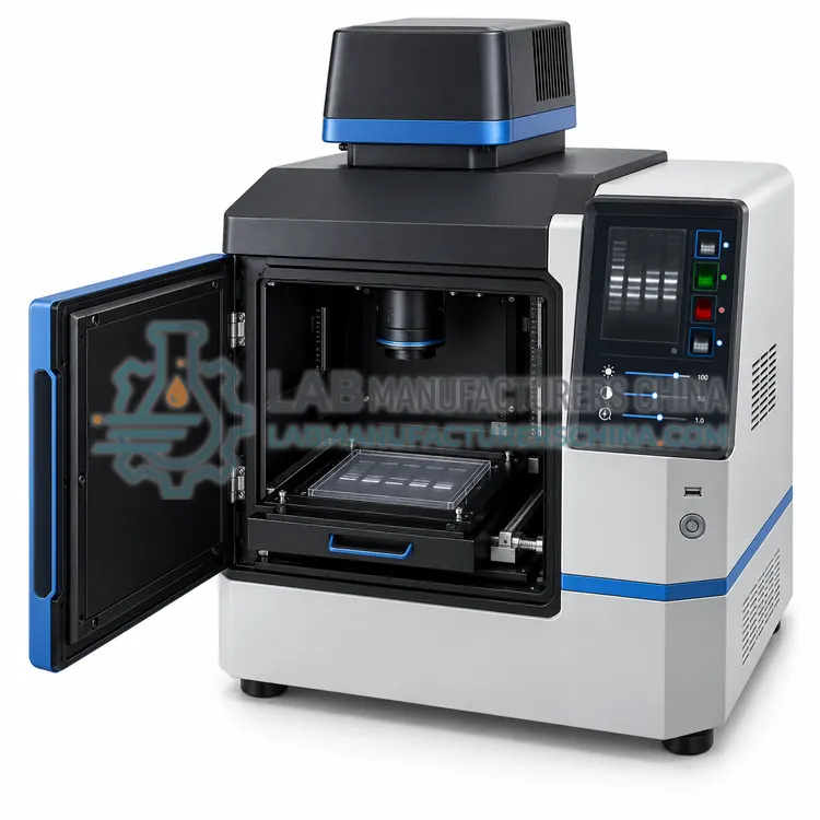
Gel documentation converts a stained or labelled gel into a digital image. A light source excites the sample, either from below through a transilluminator or from above with reflected light, and an emission filter blocks the excitation wavelengths so that only fluorescent signal reaches the camera. UV at 302 nm excites stains such as ethidium bromide, blue LEDs at about 470 nm excite SYBR-type dyes, and chemiluminescence needs no excitation because the blot emits light directly. A fixed-focus or motorized lens projects the image onto a cooled CCD or sCMOS sensor, where long exposures accumulate signal while cooling limits dark current. Software controls exposure, binning and filter selection, then displays 16-bit images and can measure band intensity, calculate molecular weights against a ladder and quantify fluorescence for densitometry.
Which Gel Documentation variants are available?
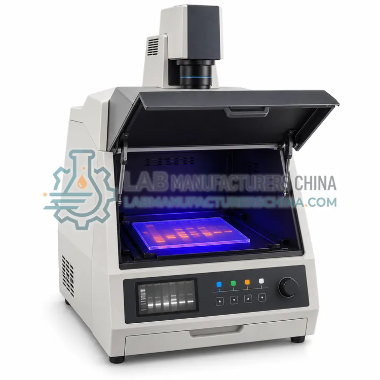
- UV Transilluminator Gel Documentation System — A darkroom imaging system with a 302 nm UV transilluminator, emission filter and cooled camera that records ethidium bromide and similar stained gels.
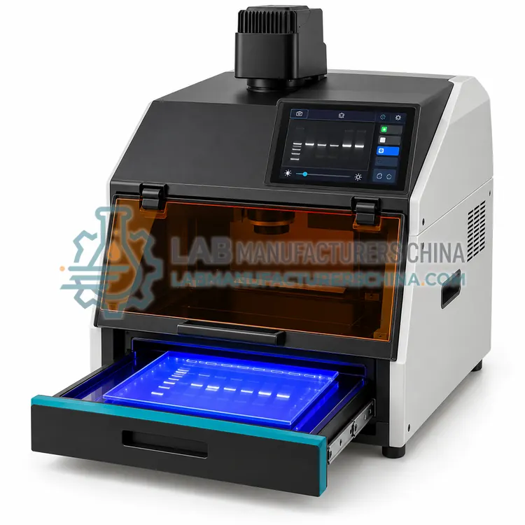
- Blue-Light LED Gel Documentation System — A documentation system with blue LED excitation near 470 nm for SYBR-type safe stains, which avoids UV hazards and reduces DNA damage.
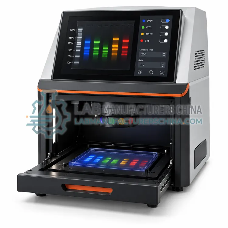
- Multifluorescence Gel Documentation System — A documentation imager with several LED excitation wavelengths and an emission filter wheel that captures multiplexed fluorescence in one gel or blot.
- Chemiluminescence Gel Documentation System — A darkroom imager with a deeply cooled camera and wide-aperture lens that records light emitted directly by chemiluminescent western blots, with no excitation source.
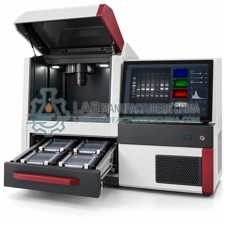
- Automated Multi-Gel Documentation System — A gel documentation platform that images several gels or blots in sequence without manual handling, using a tray magazine, automated illumination and barcode-linked file naming.
How do I choose the right variant?
Choose by the detection chemistry: UV transillumination for ethidium bromide and similar stains, blue-light LED for safer alternatives, and multifluorescence for multiplexed labels. Chemiluminescence work needs a darkroom system with a deeply cooled camera but no light source. Sensitivity depends mainly on camera quantum efficiency, cooling and lens aperture, so compare those before resolution figures. Check the excitation wavelengths and emission filters provided, whether the transilluminator is 302, 365 or dual wavelength, and how much of the gel area the lens covers. Confirm software features for band detection, ladder-based sizing, densitometry and export, and verify UV shielding and interlocks for operator safety.
Where are Gel Documentation used?
Gel Documentation are specified in agarose gel imaging after electrophoresis, western blot chemiluminescence imaging, fluorescence multiplex gel documentation, densitometric quantification of bands and preparation of publication and report figures.
Which standards and certifications apply?
Work with Gel Documentation is commonly governed by IEC 61010-1, IEC 61010-2-081, IEC 62471, IEC 61326-1 and ISO 13485 manufacturing. Instrument certifications such as CE marking are confirmed per configuration and never assumed.
What are the key considerations before purchase?
Position the system away from strong ambient light and vibration, and keep the enclosure closed during exposure. Inspect and clean the UV transilluminator filter periodically, because scratches and stains scatter light and reduce contrast. Protect the camera lens and emission filter from dust and fingerprints, storing filters in their cases. Check UV intensity with a radiometer at defined intervals, replace lamps at the rated hours and verify the interlock each time the unit is serviced. Apply a consistent exposure and aperture setting across a project so intensities are comparable, and record imaging parameters with each saved file. Ensure that users follow local rules on UV exposure, gloves and stain waste disposal.