UV Transilluminator Gel Documentation System
紫外透射凝胶成像系统
- SKU GED-0001
- Gel Documentation
- CE (to be confirmed)
- ISO 9001 manufacturing (to be confirmed)
- IEC 62471 photobiological safety report (to be confirmed)
Price on request — FOB / CIF quoted per order
Prices depend on configuration, quantity and destination port.
Key specifications
| Excitation wavelength | 302 nm standard, 254/365 nm option |
|---|---|
| Transilluminator area | 20 cm × 20 cm |
| Emission filter | 590-620 nm, filter wheel option |
| Camera type | Cooled CCD or sCMOS |
| Camera resolution | 1-16 megapixels |
| Bit depth | 16 bit |
Request a quote Download brochure · to be provided
By Lab Manufacturers China Technical TeamLast reviewed Standards: IEC 61010-1, IEC 61010-2-081, IEC 62471
What is the UV Transilluminator Gel Documentation System?
A darkroom imaging system with a 302 nm UV transilluminator, emission filter and cooled camera that records ethidium bromide and similar stained gels. It is the standard configuration for routine agarose gel documentation.
What is the UV Transilluminator Gel Documentation System?
A darkroom imaging system with a 302 nm UV transilluminator, emission filter and cooled camera that records ethidium bromide and similar stained gels. It is the standard configuration for routine agarose gel documentation.
Overview
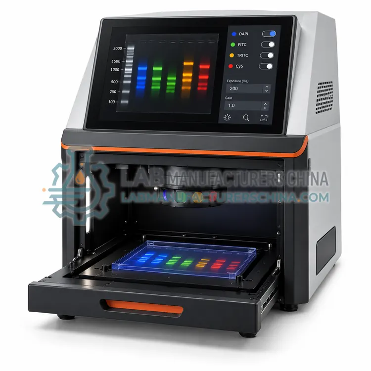
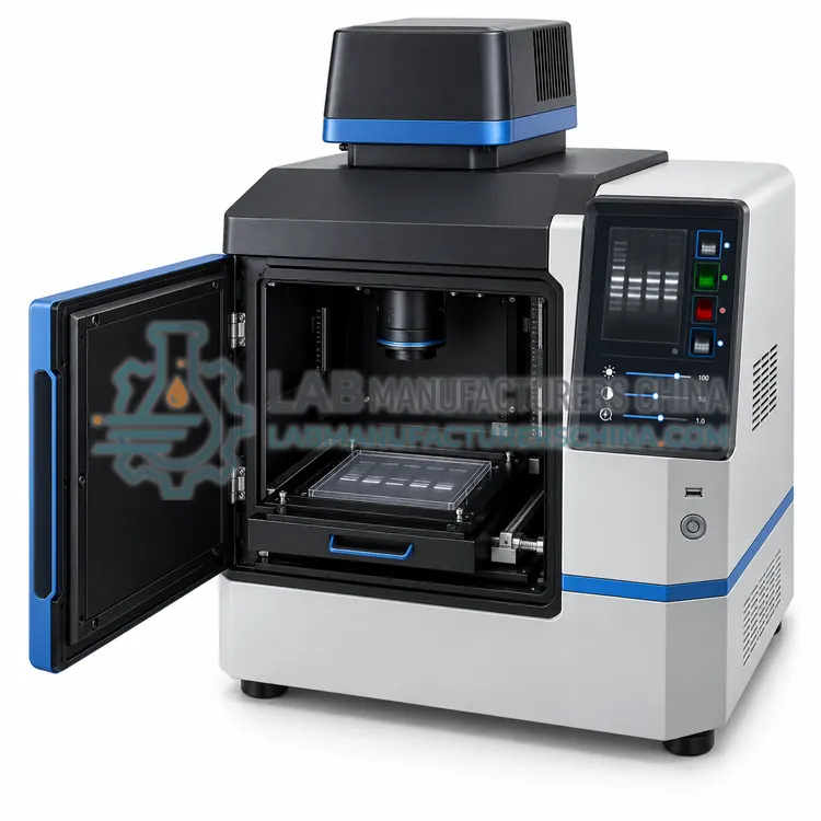
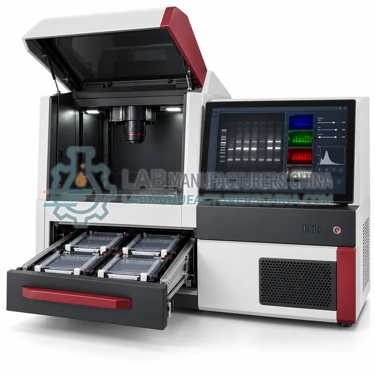
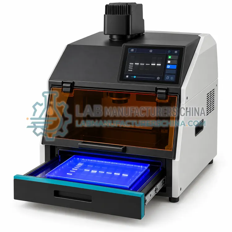
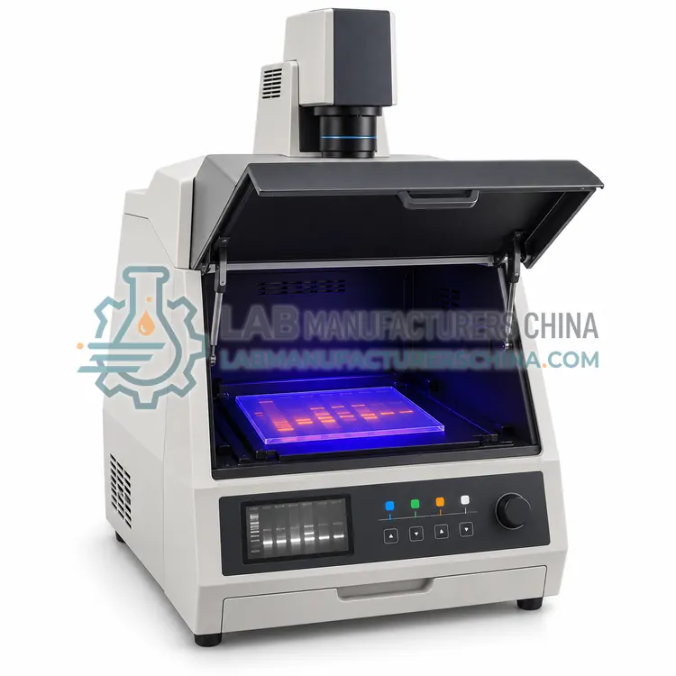
The UV gel documentation system places the gel on a transilluminator inside a light-tight hood lined with UV-absorbing material. A 302 nm lamp or a dual 254 and 365 nm option excites the intercalated stain, and an emission filter, typically 590 to 620 nm for ethidium bromide, blocks the excitation light. The camera is a cooled CCD or sCMOS with 1 to 16 megapixels and 16-bit output, and most systems use a motorized zoom lens with f/1.2 to f/1.4 aperture for framing. Software sets exposure, gain and binning, captures the image, overlays a grid and can compute molecular weights from a ladder band set. UV shielding, a door interlock and often an amber viewing window protect the operator, and some models add white epi-illumination for colour-marker images.
How does it work?
Gel documentation converts a stained or labelled gel into a digital image. A light source excites the sample, either from below through a transilluminator or from above with reflected light, and an emission filter blocks the excitation wavelengths so that only fluorescent signal reaches the camera. UV at 302 nm excites stains such as ethidium bromide, blue LEDs at about 470 nm excite SYBR-type dyes, and chemiluminescence needs no excitation because the blot emits light directly. A fixed-focus or motorized lens projects the image onto a cooled CCD or sCMOS sensor, where long exposures accumulate signal while cooling limits dark current. Software controls exposure, binning and filter selection, then displays 16-bit images and can measure band intensity, calculate molecular weights against a ladder and quantify fluorescence for densitometry.
What are the key features?
- 302 nm UV transilluminator with optional 254 or 365 nm.
- Emission filter matched to ethidium bromide and similar stains.
- Cooled CCD or sCMOS camera with 16-bit output.
- Motorized zoom lens with wide aperture for short exposures.
- Software with ladder-based sizing and densitometry.
- Door interlock and UV-absorbing enclosure.
What are the technical specifications?
A typical uv transilluminator gel documentation system configuration offers excitation wavelength of 302 nm standard, 254/365 nm option, transilluminator area of 20 cm × 20 cm, emission filter of 590-620 nm, filter wheel option, camera type of Cooled CCD or sCMOS, camera resolution of 1-16 megapixels and bit depth of 16 bit. Values are indicative of the product class; the exact figures for each configuration are confirmed on the quotation and the calibration documents supplied with the instrument.
Which applications is it used for?
The UV Transilluminator Gel Documentation System is used in agarose gel documentation, restriction digest and PCR product images, densitometric band quantification and teaching laboratory gel records. Across the wider Gel Documentation class, laboratories also deploy it for agarose gel imaging after electrophoresis, western blot chemiluminescence imaging and fluorescence multiplex gel documentation.
Which standards and certifications apply?
Relevant standards and methods include IEC 61010-1, IEC 61010-2-081, IEC 62471, IEC 61326-1 and ISO 13485 manufacturing. Certification candidates for this model are CE, ISO 9001 manufacturing and IEC 62471 photobiological safety report; each is to be confirmed for the specific configuration before shipment, and certificates are supplied only where they have been issued.
Position the system away from strong ambient light and vibration, and keep the enclosure closed during exposure. Inspect and clean the UV transilluminator filter periodically, because scratches and stains scatter light and reduce contrast. Protect the camera lens and emission filter from dust and fingerprints, storing filters in their cases. Check UV intensity with a radiometer at defined intervals, replace lamps at the rated hours and verify the interlock each time the unit is serviced. Apply a consistent exposure and aperture setting across a project so intensities are comparable, and record imaging parameters with each saved file. Ensure that users follow local rules on UV exposure, gloves and stain waste disposal.
How do I select the right UV Transilluminator Gel Documentation System?
Select this configuration for routine DNA gels stained with ethidium bromide or equivalent dyes. Compare camera cooling and quantum efficiency against your weakest expected signal, verify the transilluminator size covers your largest gel, and check whether a dual-wavelength option is needed. Confirm that the analysis software includes sizing and densitometry without extra licence cost.
How does it compare with other Gel Documentation variants?
Choose by the detection chemistry: UV transillumination for ethidium bromide and similar stains, blue-light LED for safer alternatives, and multifluorescence for multiplexed labels. Chemiluminescence work needs a darkroom system with a deeply cooled camera but no light source. Sensitivity depends mainly on camera quantum efficiency, cooling and lens aperture, so compare those before resolution figures. Check the excitation wavelengths and emission filters provided, whether the transilluminator is 302, 365 or dual wavelength, and how much of the gel area the lens covers. Confirm software features for band detection, ladder-based sizing, densitometry and export, and verify UV shielding and interlocks for operator safety.
Full specifications
| Excitation wavelength | 302 nm standard, 254/365 nm option |
|---|---|
| Transilluminator area | 20 cm × 20 cm |
| Emission filter | 590-620 nm, filter wheel option |
| Camera type | Cooled CCD or sCMOS |
| Camera resolution | 1-16 megapixels |
| Bit depth | 16 bit |
| Lens | Motorized zoom, f/1.2-1.4 |
| Exposure range | 0.1 s to 60 s |
| Software | Capture, sizing, densitometry, 16-bit TIFF export |
| Safety | UV shield, door interlock, amber window |
Indicative values for this product class. Final specifications are confirmed on the quotation and in the calibration documents supplied with the instrument.
Applications
- Agarose gel documentation
- Restriction digest and PCR product images
- Densitometric band quantification
- Teaching laboratory gel records
Key features
- 302 nm UV transilluminator with optional 254 or 365 nm.
- Emission filter matched to ethidium bromide and similar stains.
- Cooled CCD or sCMOS camera with 16-bit output.
- Motorized zoom lens with wide aperture for short exposures.
- Software with ladder-based sizing and densitometry.
- Door interlock and UV-absorbing enclosure.
Frequently asked questions about UV Transilluminator Gel Documentation System
302 nm gives stronger signal from ethidium bromide and most intercalating stains, while 365 nm causes less DNA damage for downstream applications such as cloning. Dual-wavelength models let you switch between the two.
Short exposure at 302 nm during imaging causes some nicking, so excise bands quickly and use 365 nm or blue light when the fragment will be cloned or sequenced after extraction.
Only if white epi-illumination is fitted and the filter wheel includes a clear position. Standard UV systems image fluorescent signals, so confirm the illumination options if you also run Coomassie or silver-stained gels.
UV at 302 nm excites ethidium bromide and similar intercalating stains, blue LEDs near 470 nm excite SYBR-type dyes, and white or coloured epi-illumination suits colorimetric stains. Chemiluminescence needs no light source at all.
Moderately cooled cameras are adequate for bright UV-excited gels, while chemiluminescence and weak fluorescence need cooling to -30 °C or below to keep dark current low during long exposures. Cooling extends usable exposure time.
Only with care. Intensity depends on exposure, aperture, stain concentration, gel thickness and illumination uniformity. Use fixed settings, include a ladder or standard on each gel, and confirm that no band is saturated before comparing samples.
Related models