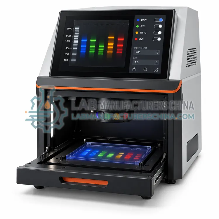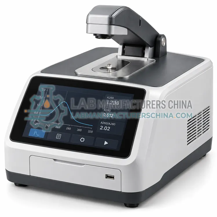
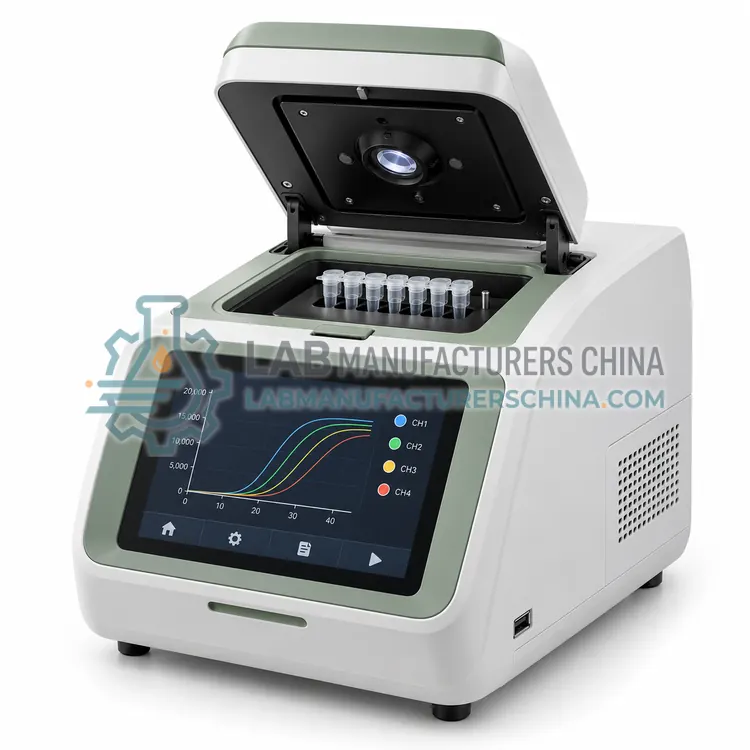
Fluorescence Nucleic Acid Quantification System
荧光核酸定量系统
- SKU NUC-0002
- Nucleic Acid Analysis
- CE (to be confirmed)
- ISO 9001 manufacturing (to be confirmed)
- IEC 61010-1 test report (to be confirmed)
Price on request — FOB / CIF quoted per order
Prices depend on configuration, quantity and destination port.
Key specifications
| Excitation wavelength | 470-490 nm |
|---|---|
| Emission wavelength | 510-530 nm |
| Detection limit | 0.2 ng/mL with high-sensitivity kit |
| Linear range | 0.2-1,000 ng/mL, extended with small-volume mode |
| Sample volume | 1-20 µL |
| Calibration | 2-5 point standard curve per kit |
Request a quote Download brochure · to be provided
By Lab Manufacturers China Technical TeamLast reviewed Standards: ISO 20395, ISO 13485 manufacturing, MIQE guidelines
What is the Fluorescence Nucleic Acid Quantification System?
A fluorometer that uses dyes specific for double-stranded DNA or RNA to quantify as little as a few picograms in 1 to 20 µL samples. It tolerates protein, salt and free nucleotides that distort absorbance readings.
What is the Fluorescence Nucleic Acid Quantification System?
A fluorometer that uses dyes specific for double-stranded DNA or RNA to quantify as little as a few picograms in 1 to 20 µL samples. It tolerates protein, salt and free nucleotides that distort absorbance readings.
Overview
The fluorescence quantification system uses a selective dye chemistry, for example PicoGreen for double-stranded DNA, RiboGreen for RNA or a broad dye for single-stranded DNA. A standard curve of two to five points prepared with the same kit converts fluorescence into concentration, and each assay kit defines its own linear range, commonly 0.2 to 1,000 ng/mL for high-sensitivity kits and up to 1,000 ng/µL in a small-volume format. Excitation and emission filters are matched to the dye, usually blue excitation around 480 nm with green emission near 520 nm, and sample volume can be as low as 1 µL with a dedicated adapter. Because the dye reports only the target, results agree closely between crude lysates and purified samples, which improves normalisation for sequencing and qPCR.
How does it work?
Two measurement principles dominate. Absorbance analysis uses a 1 mm or 0.5 mm pathlength in a microvolume cell and reads at 260 nm, where nucleic acids absorb strongly, then scales the result to a standard 10 mm path. A260 of 1.0 corresponds to about 50 µg/mL double-stranded DNA, 33 µg/mL single-stranded DNA and 40 µg/mL RNA, and ratios at 260/280 and 260/230 indicate protein or salt contamination. Fluorescence methods use dyes that bind specifically to double-stranded DNA or to total RNA, so signal comes only from the target, which makes them more tolerant of protein, salts and free nucleotides. Capillary electrophoresis separates fragments in a sieving polymer and reports sizes in base pairs together with molarity. Automated workstations combine measurement with pipetting so samples can be diluted to a target concentration.
What are the key features?
- Dye chemistry selective for double-stranded DNA or RNA.
- Detection down to a few picograms per assay.
- Two to five point standard curve with kit-specific controls.
- Sample volume from 1 µL with adapter.
- Excitation and emission filters matched to common dyes.
- Lower sensitivity to protein, salt and nucleotide contamination.
What are the technical specifications?
A typical fluorescence nucleic acid quantification system configuration offers excitation wavelength of 470-490 nm, emission wavelength of 510-530 nm, detection limit of 0.2 ng/mL with high-sensitivity kit, linear range of 0.2-1,000 ng/mL, extended with small-volume mode, sample volume of 1-20 µL and calibration of 2-5 point standard curve per kit. Values are indicative of the product class; the exact figures for each configuration are confirmed on the quotation and the calibration documents supplied with the instrument.
Which applications is it used for?
The Fluorescence Nucleic Acid Quantification System is used in sequencing library quantification, low-yield DNA or RNA sample measurement, template normalisation before qPCR and quality control of extracted nucleic acids. Across the wider Nucleic Acid Analysis class, laboratories also deploy it for next-generation sequencing library quality control, template quantification before qPCR and plasmid and oligonucleotide quality checks.
Which standards and certifications apply?
Relevant standards and methods include ISO 20395, ISO 13485 manufacturing, MIQE guidelines, CLSI MM06-A2, IEC 61010-1 and IVDR (EU) 2017/746. Certification candidates for this model are CE, ISO 9001 manufacturing and IEC 61010-1 test report; each is to be confirmed for the specific configuration before shipment, and certificates are supplied only where they have been issued.
Blank the instrument with the same buffer as the sample, because buffer absorbance differs and elution buffers containing guanidine or EDTA give misleading ratios. Keep the measurement pedestal clean and wipe it between samples to avoid carryover, and check microvolume accuracy with a reference standard periodically. Fluorescent assays need fresh standards and protection of dye reagents from light, and dye-based concentration depends on assay chemistry, so do not compare results from different kits. For fragment analysis, follow the vendor's sample and ladder preparation closely, since ionic strength affects injection. Store eluted nucleic acids cold, avoid repeated freeze-thaw, and record concentration, ratios and method with every sample.
How do I select the right Fluorescence Nucleic Acid Quantification System?
Select the fluorometer when samples are dilute, contaminated or when only double-stranded DNA should be counted, as with sequencing libraries. Confirm that the excitation and emission filters match the dye kits you use, and check the low and high ends of the linear range against your expected concentrations. Include kit consumption in the running cost estimate.
How does it compare with other Nucleic Acid Analysis variants?
Choose the method by how much selectivity you need. Absorbance is fast, inexpensive and sufficient for routine plasmid or PCR checks, but it cannot distinguish DNA from RNA and is affected by protein, phenol and salts. Fluorescence dyes are selective for double-stranded DNA or RNA and need as little as a few picograms, which suits sequencing libraries and low-yield samples. Capillary electrophoresis adds size and integrity information that no spectrophotometer provides, which matters for library insert size and RNA integrity. Throughput then decides between single-tube devices, microplate readers and automated workstations that combine measurement with normalisation. Check the minimum sample volume, detection limit, linear range, required standards and the export format your data system accepts.
Full specifications
| Excitation wavelength | 470-490 nm |
|---|---|
| Emission wavelength | 510-530 nm |
| Detection limit | 0.2 ng/mL with high-sensitivity kit |
| Linear range | 0.2-1,000 ng/mL, extended with small-volume mode |
| Sample volume | 1-20 µL |
| Calibration | 2-5 point standard curve per kit |
| Assay time | 1-5 min including incubation |
| Dye compatibility | dsDNA, RNA and ssDNA assay kits |
| Data output | Concentration, standard curve, raw fluorescence |
| Power supply | 100-240 V, 50/60 Hz, 30-60 W |
Indicative values for this product class. Final specifications are confirmed on the quotation and in the calibration documents supplied with the instrument.
Applications
- Sequencing library quantification
- Low-yield DNA or RNA sample measurement
- Template normalisation before qPCR
- Quality control of extracted nucleic acids
Key features
- Dye chemistry selective for double-stranded DNA or RNA.
- Detection down to a few picograms per assay.
- Two to five point standard curve with kit-specific controls.
- Sample volume from 1 µL with adapter.
- Excitation and emission filters matched to common dyes.
- Lower sensitivity to protein, salt and nucleotide contamination.
Frequently asked questions about Fluorescence Nucleic Acid Quantification System
Absorbance measures everything that absorbs at 260 nm, including RNA, free nucleotides and some proteins, while a double-stranded dye measures only the target. A large gap usually means contamination or degradation in the sample.
Prepare a fresh standard curve for each working session or at least each day, since dye activity and instrument response drift. Use the standards supplied with the kit, because other DNA sources may behave differently in the assay.
High concentrations can inhibit dye binding, so dilute samples into the kit buffer as instructed. Most kits tolerate typical elution buffers, but validating with your own buffer before routine use is recommended.
A260/280 near 1.8 for DNA and 2.0 for RNA indicates low protein contamination, while A260/230 above about 2.0 suggests little carryover of salts, phenols or guanidine. Values well below these ranges usually mean the sample needs repurification.
Use fluorescence when the sample is scarce, when the template contains protein, phenol or free nucleotides, or when only double-stranded DNA should be measured. Dyes such as PicoGreen or RiboGreen report the target specifically, so values are less affected by contaminants.
For sequencing libraries, RNA samples and amplicon panels, yes. Concentration alone cannot show whether a library insert is the correct length or whether RNA is degraded, and those properties determine sequencing and qPCR performance.
Related models
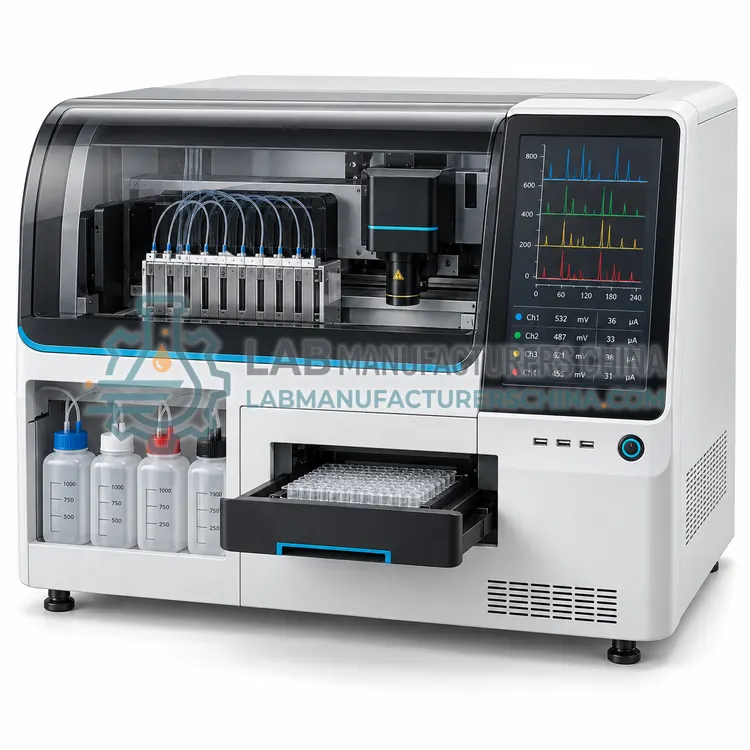
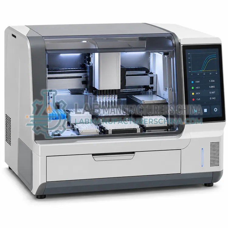
Automated Nucleic Acid Quantification and Normalization Workstation
自动核酸定量与均一化工作站
- CE (to be confirmed)
- +2