Chemiluminescence Gel Documentation System
化学发光凝胶成像系统
- SKU GED-0004
- Gel Documentation
- CE (to be confirmed)
- ISO 9001 manufacturing (to be confirmed)
- IEC 61010-1 test report (to be confirmed)
Price on request — FOB / CIF quoted per order
Prices depend on configuration, quantity and destination port.
Key specifications
| Detection mode | Chemiluminescence, no excitation |
|---|---|
| Camera type | Cooled CCD or sCMOS |
| Camera cooling | -30 to -60 °C |
| Bit depth | 16 bit |
| Lens aperture | f/0.95-1.4 fixed or motorized |
| Exposure range | 1 s to 30 min |
Request a quote Download brochure · to be provided
By Lab Manufacturers China Technical TeamLast reviewed Standards: IEC 61010-1, IEC 61010-2-081, IEC 62471
What is the Chemiluminescence Gel Documentation System?
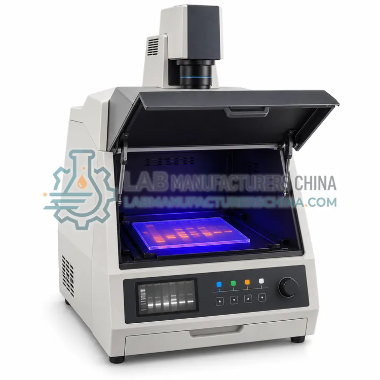
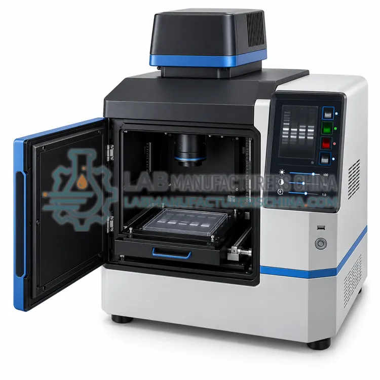
A darkroom imager with a deeply cooled camera and wide-aperture lens that records light emitted directly by chemiluminescent western blots, with no excitation source. It is chosen for sensitive detection of low-abundance proteins.
What is the Chemiluminescence Gel Documentation System?
A darkroom imager with a deeply cooled camera and wide-aperture lens that records light emitted directly by chemiluminescent western blots, with no excitation source. It is chosen for sensitive detection of low-abundance proteins.
Overview
The chemiluminescence system has no illumination optics because the blot emits light from an enzymatic reaction, usually horseradish peroxidase with a luminol substrate. Sensitivity therefore depends on collecting as much light as possible, which means a lens with an aperture of f/0.95 to f/1.4, a camera cooled to -30 to -60 °C to reduce dark current, and exposures from seconds to 30 minutes. Pixel binning increases sensitivity at the cost of resolution, and software can capture a series of exposures so that both weak and strong bands are within the linear range. Some instruments add colourimetric and fluorescence modes with LED or UV light sources, making them multi-purpose. Careful substrate application, consistent timing between blots and an even membrane surface are essential for reproducible quantitation.
How does it work?
Gel documentation converts a stained or labelled gel into a digital image. A light source excites the sample, either from below through a transilluminator or from above with reflected light, and an emission filter blocks the excitation wavelengths so that only fluorescent signal reaches the camera. UV at 302 nm excites stains such as ethidium bromide, blue LEDs at about 470 nm excite SYBR-type dyes, and chemiluminescence needs no excitation because the blot emits light directly. A fixed-focus or motorized lens projects the image onto a cooled CCD or sCMOS sensor, where long exposures accumulate signal while cooling limits dark current. Software controls exposure, binning and filter selection, then displays 16-bit images and can measure band intensity, calculate molecular weights against a ladder and quantify fluorescence for densitometry.
What are the key features?
- No excitation source needed, so only emitted light is imaged.
- Deeply cooled camera at -30 to -60 °C for low dark current.
- Wide-aperture lens f/0.95 to f/1.4 for light collection.
- Exposure series from seconds to 30 minutes.
- Pixel binning option to increase sensitivity.
- Optional colourimetric and fluorescence imaging modes.
What are the technical specifications?
A typical chemiluminescence gel documentation system configuration offers detection mode of Chemiluminescence, no excitation, camera type of Cooled CCD or sCMOS, camera cooling of -30 to -60 °C, bit depth of 16 bit, lens aperture of f/0.95-1.4 fixed or motorized and exposure range of 1 s to 30 min. Values are indicative of the product class; the exact figures for each configuration are confirmed on the quotation and the calibration documents supplied with the instrument.
Which applications is it used for?
The Chemiluminescence Gel Documentation System is used in western blot chemiluminescence detection, protein expression comparison, immunoprecipitation blot analysis and sensitive detection of low-abundance proteins. Across the wider Gel Documentation class, laboratories also deploy it for agarose gel imaging after electrophoresis, western blot chemiluminescence imaging and fluorescence multiplex gel documentation.
Which standards and certifications apply?
Relevant standards and methods include IEC 61010-1, IEC 61010-2-081, IEC 62471, IEC 61326-1 and ISO 13485 manufacturing. Certification candidates for this model are CE, ISO 9001 manufacturing and IEC 61010-1 test report; each is to be confirmed for the specific configuration before shipment, and certificates are supplied only where they have been issued.
Position the system away from strong ambient light and vibration, and keep the enclosure closed during exposure. Inspect and clean the UV transilluminator filter periodically, because scratches and stains scatter light and reduce contrast. Protect the camera lens and emission filter from dust and fingerprints, storing filters in their cases. Check UV intensity with a radiometer at defined intervals, replace lamps at the rated hours and verify the interlock each time the unit is serviced. Apply a consistent exposure and aperture setting across a project so intensities are comparable, and record imaging parameters with each saved file. Ensure that users follow local rules on UV exposure, gloves and stain waste disposal.
How do I select the right Chemiluminescence Gel Documentation System?
Select this variant for chemiluminescent blots, particularly when target proteins are of low abundance. Compare camera cooling, quantum efficiency, lens aperture and binning options, as these set the detection limit. Check whether the software warns about saturation and can capture exposure series, and confirm that the darkroom is fully light-tight for long exposures.
How does it compare with other Gel Documentation variants?
Choose by the detection chemistry: UV transillumination for ethidium bromide and similar stains, blue-light LED for safer alternatives, and multifluorescence for multiplexed labels. Chemiluminescence work needs a darkroom system with a deeply cooled camera but no light source. Sensitivity depends mainly on camera quantum efficiency, cooling and lens aperture, so compare those before resolution figures. Check the excitation wavelengths and emission filters provided, whether the transilluminator is 302, 365 or dual wavelength, and how much of the gel area the lens covers. Confirm software features for band detection, ladder-based sizing, densitometry and export, and verify UV shielding and interlocks for operator safety.
Full specifications
| Detection mode | Chemiluminescence, no excitation |
|---|---|
| Camera type | Cooled CCD or sCMOS |
| Camera cooling | -30 to -60 °C |
| Bit depth | 16 bit |
| Lens aperture | f/0.95-1.4 fixed or motorized |
| Exposure range | 1 s to 30 min |
| Binning | 1 × 1 to 8 × 8 |
| Imaging area | Up to 20 cm × 25 cm |
| Additional modes | Optional colorimetric and fluorescence |
| Software | Exposure series, saturation warning, quantitation |
Indicative values for this product class. Final specifications are confirmed on the quotation and in the calibration documents supplied with the instrument.
Applications
- Western blot chemiluminescence detection
- Protein expression comparison
- Immunoprecipitation blot analysis
- Sensitive detection of low-abundance proteins
Key features
- No excitation source needed, so only emitted light is imaged.
- Deeply cooled camera at -30 to -60 °C for low dark current.
- Wide-aperture lens f/0.95 to f/1.4 for light collection.
- Exposure series from seconds to 30 minutes.
- Pixel binning option to increase sensitivity.
- Optional colourimetric and fluorescence imaging modes.
Frequently asked questions about Chemiluminescence Gel Documentation System
A chemiluminescent blot emits very little light, and the amount captured scales with lens aperture area. An f/0.95 lens collects several times more light than an f/1.4 lens, shortening exposure and reducing noise.
Binning combines neighbouring pixels and increases sensitivity at the cost of spatial resolution. It helps for very weak bands, but avoid it when fine band separation matters, since resolution may fall below what quantitation requires.
Uneven substrate coverage, variable timing between substrate addition and imaging, membrane edges drying and saturated bands. Standardise each step, image a series of exposures and confirm that measured bands are below saturation.
UV at 302 nm excites ethidium bromide and similar intercalating stains, blue LEDs near 470 nm excite SYBR-type dyes, and white or coloured epi-illumination suits colorimetric stains. Chemiluminescence needs no light source at all.
Moderately cooled cameras are adequate for bright UV-excited gels, while chemiluminescence and weak fluorescence need cooling to -30 °C or below to keep dark current low during long exposures. Cooling extends usable exposure time.
Only with care. Intensity depends on exposure, aperture, stain concentration, gel thickness and illumination uniformity. Use fixed settings, include a ladder or standard on each gel, and confirm that no band is saturated before comparing samples.
Related models
Automated Nucleic Acid Quantification and Normalization Workstation
自动核酸定量与均一化工作站
- CE (to be confirmed)
- +2