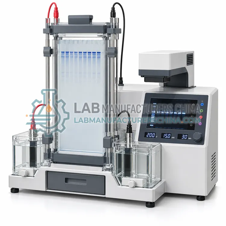
How do Electrophoresis work?
In electrophoresis, an electric field drives charged molecules through a gel matrix: nucleic acids carry a negative charge from their phosphate backbone and migrate toward the anode, while their size and conformation determine how quickly they pass through the pores. Agarose gels of 0.5 to 2 percent resolve fragments from about 100 bp to 20 kb, and a buffer such as TAE or TBE carries the current between electrodes. Voltage is set per centimetre of gel length, commonly 5 to 10 V/cm, because higher voltages accelerate runs but heat the gel and blur bands. Sample loading dye adds density and tracking dyes, and a size marker gives fragment lengths by comparison. Detection uses intercalating stains under UV or blue light, or fluorescent labels in chip systems. Denaturing urea-PAGE resolves single-base differences in short oligonucleotides.
Which Electrophoresis variants are available?
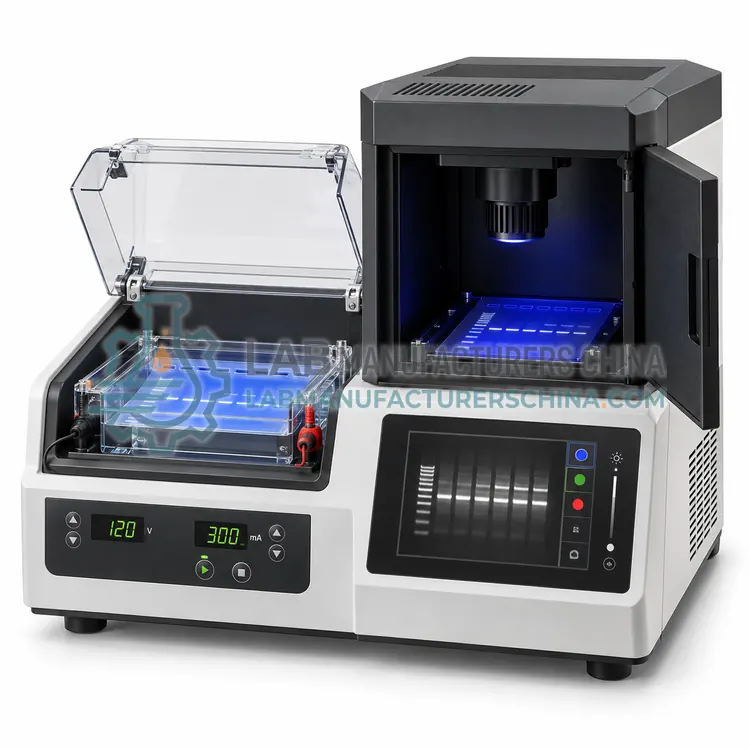
- Automated Agarose Gel Electrophoresis System with On-Board Imaging — An integrated instrument that loads precast agarose gels, runs the separation and captures an image without user handling of the gel.
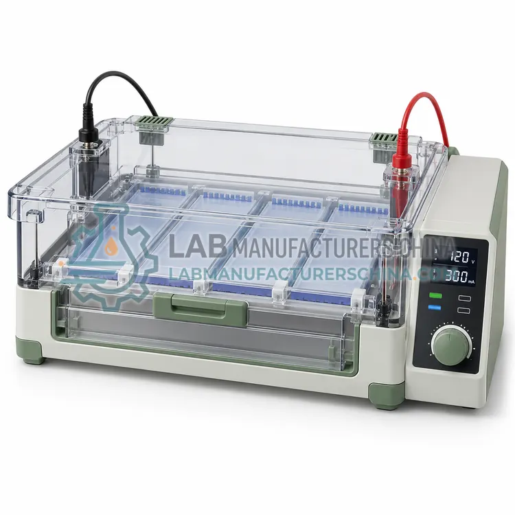
- Multi-Channel Agarose Gel Electrophoresis System — A horizontal tank with multiple independent lanes, trays or sub-cells, letting several users or protocols run at the same time.
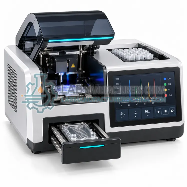
- Microfluidic Chip Electrophoresis System — A benchtop analyser that separates fluorescently labelled nucleic acid fragments inside a microfluidic chip and reports digital sizes and concentrations.
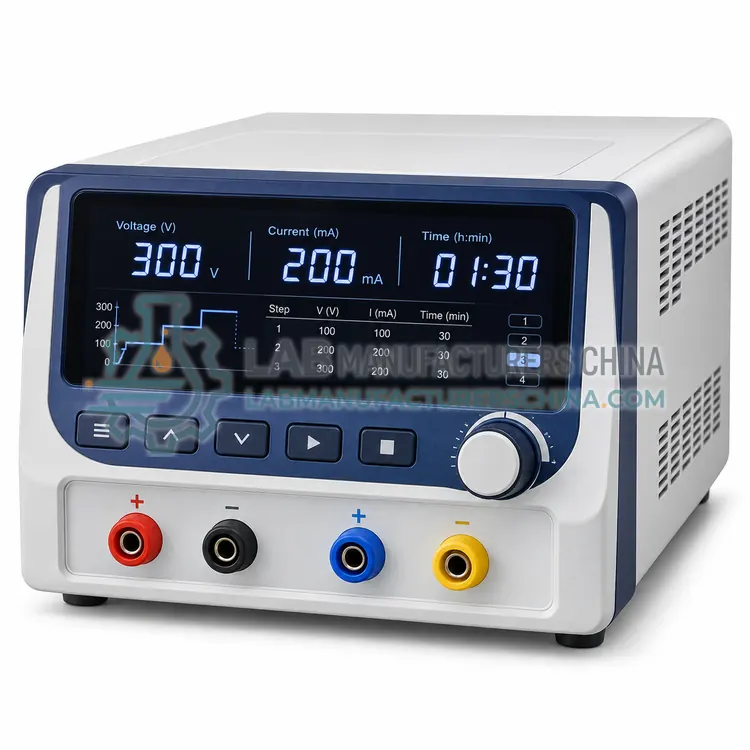
- Programmable Electrophoresis Power Supply — A regulated power supply that delivers constant voltage, current or power to one or more electrophoresis gel tanks, with timed programs and safety limits.
- Denaturing Urea-PAGE System for Oligonucleotide Analysis — A vertical electrophoresis system with heated plates and urea-denaturing gels that resolves short single-stranded nucleic acids differing by one base.
How do I choose the right variant?
Match the method to the molecule and the resolution needed. Agarose in a horizontal tank is standard for double-stranded DNA fragments and PCR products, denaturing urea-PAGE for oligonucleotides and single-stranded fragments, and sieving-polymer chip systems for accurate sizing and quantification of many samples. Throughput then decides between manual tanks, multi-channel units and automated systems that combine running and imaging. Consider gel format and well count, buffer volume and cooling, and the detection method, because stains, UV exposure and fluorescence optics differ in sensitivity and safety. For power supplies, check voltage, current and power limits, programmability, and whether constant voltage, constant current or constant power modes are available.
Where are Electrophoresis used?
Electrophoresis are specified in PCR product verification, DNA fragment sizing and restriction mapping, next-generation sequencing library quality control, oligonucleotide purity analysis and teaching and method development.
Which standards and certifications apply?
Work with Electrophoresis is commonly governed by IEC 61010-1, IEC 61010-2-081, IEC 61326-1, ISO 13485 manufacturing and CLSI MM13-A. Instrument certifications such as CE marking are confirmed per configuration and never assumed.
What are the key considerations before purchase?
Prepare gels in a dedicated area and pour them on a level surface. Use fresh buffer at the recommended dilution, since reused buffer changes pH and conductivity, and check that electrode wires and connectors are clean. Keep the tank covered during runs to limit evaporation and protect operators from UV or electrical hazards, and never touch buffer or electrodes while a run is active. Choose low-toxicity stains where possible and follow local disposal rules for ethidium bromide or other intercalating agents. Clean and rinse tanks after each run to remove residual stain and agarose. Verify power supply calibration and check for cracked gel trays, worn leads and loose lid interlocks at fixed intervals.