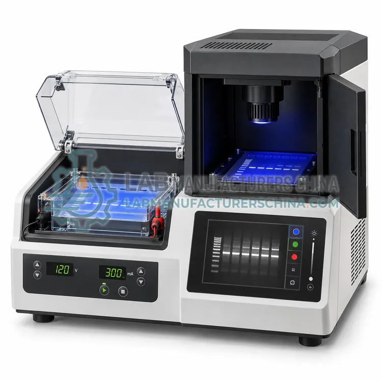
Automated Agarose Gel Electrophoresis System with On-Board Imaging
带机上成像的自动琼脂糖凝胶电泳系统
- CE (to be confirmed)
- +2
Welcome to Lab Manufacturers China — laboratory instruments direct from a Chinese manufacturer
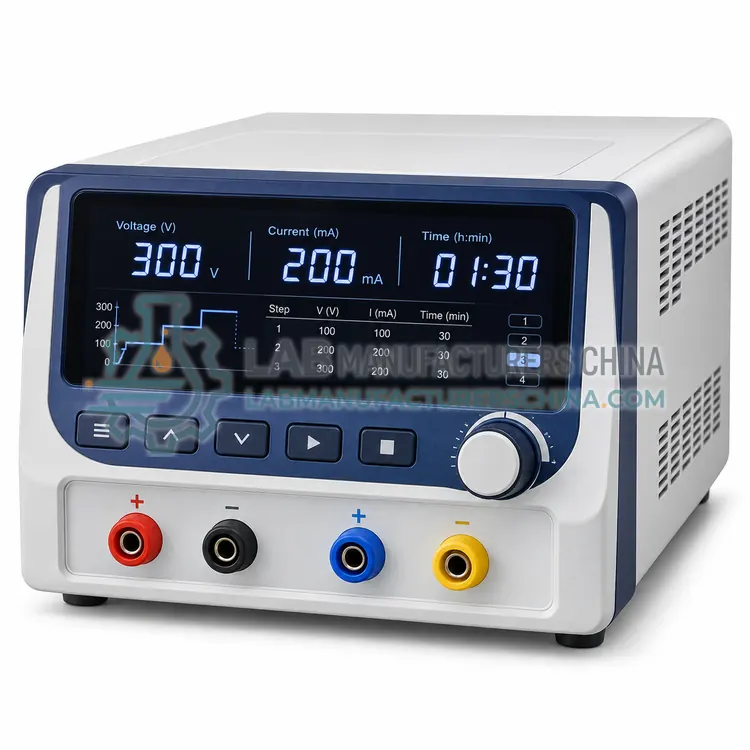
可编程电泳电源
Price on request — FOB / CIF quoted per order
Prices depend on configuration, quantity and destination port.
| Maximum voltage | 200-5,000 V depending on model |
|---|---|
| Maximum current | 200-2,000 mA |
| Maximum power | 100-400 W |
| Control modes | Constant voltage, current or power |
| Programs stored | 10-100 multi-step methods |
| Timer range | 1 min to 99 h, or volt-hour mode |
By Lab Manufacturers China Technical TeamLast reviewed Standards: IEC 61010-1, IEC 61010-2-081, IEC 61326-1
A regulated power supply that delivers constant voltage, current or power to one or more electrophoresis gel tanks, with timed programs and safety limits. It is the component that largely determines run reproducibility.
A regulated power supply that delivers constant voltage, current or power to one or more electrophoresis gel tanks, with timed programs and safety limits. It is the component that largely determines run reproducibility.
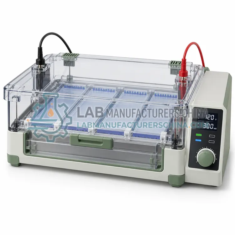
The electrophoresis power supply converts mains electricity into controlled direct current for the buffer chambers. Output ratings range from 200 V and 500 mA for small horizontal tanks to 3,000 to 5,000 V and 200 mA for sequencing and isoelectric focusing applications. Users set a limit for voltage, current and power, and the supply holds one parameter constant while the others vary with buffer conductance and temperature. Programmable models store multi-step methods, for example a low-voltage sample entry step followed by a higher-voltage separation, and can run timed or volt-hour protocols. Safety features include no-load and short-circuit detection, arc protection, earth-leakage detection and lid interlocks that cut output when a tank is opened. Multiple outlets allow two or four tanks to run at once.
In electrophoresis, an electric field drives charged molecules through a gel matrix: nucleic acids carry a negative charge from their phosphate backbone and migrate toward the anode, while their size and conformation determine how quickly they pass through the pores. Agarose gels of 0.5 to 2 percent resolve fragments from about 100 bp to 20 kb, and a buffer such as TAE or TBE carries the current between electrodes. Voltage is set per centimetre of gel length, commonly 5 to 10 V/cm, because higher voltages accelerate runs but heat the gel and blur bands. Sample loading dye adds density and tracking dyes, and a size marker gives fragment lengths by comparison. Detection uses intercalating stains under UV or blue light, or fluorescent labels in chip systems. Denaturing urea-PAGE resolves single-base differences in short oligonucleotides.
A typical programmable electrophoresis power supply configuration offers maximum voltage of 200-5,000 V depending on model, maximum current of 200-2,000 mA, maximum power of 100-400 W, control modes of Constant voltage, current or power, programs stored of 10-100 multi-step methods and timer range of 1 min to 99 h, or volt-hour mode. Values are indicative of the product class; the exact figures for each configuration are confirmed on the quotation and the calibration documents supplied with the instrument.
The Programmable Electrophoresis Power Supply is used in driving horizontal and vertical gel tanks, SDS-PAGE and native protein gels, isoelectric focusing and 2D-PAGE and long-duration nucleic acid separations. Across the wider Electrophoresis class, laboratories also deploy it for PCR product verification, DNA fragment sizing and restriction mapping and next-generation sequencing library quality control.
Relevant standards and methods include IEC 61010-1, IEC 61010-2-081, IEC 61326-1, ISO 13485 manufacturing and CLSI MM13-A. Certification candidates for this model are CE, ISO 9001 manufacturing and IEC 61010-1 test report; each is to be confirmed for the specific configuration before shipment, and certificates are supplied only where they have been issued.
Prepare gels in a dedicated area and pour them on a level surface. Use fresh buffer at the recommended dilution, since reused buffer changes pH and conductivity, and check that electrode wires and connectors are clean. Keep the tank covered during runs to limit evaporation and protect operators from UV or electrical hazards, and never touch buffer or electrodes while a run is active. Choose low-toxicity stains where possible and follow local disposal rules for ethidium bromide or other intercalating agents. Clean and rinse tanks after each run to remove residual stain and agarose. Verify power supply calibration and check for cracked gel trays, worn leads and loose lid interlocks at fixed intervals.
Select the power supply by the maximum voltage and current your protocols need, with headroom of about 20 percent, and by the number of tanks you run at once. Choose constant-voltage operation for reproducible fragment positions and programmable multi-step methods when sample entry and separation need different settings. Confirm the safety features, especially earth-leakage detection and lid interlock compatibility.
Match the method to the molecule and the resolution needed. Agarose in a horizontal tank is standard for double-stranded DNA fragments and PCR products, denaturing urea-PAGE for oligonucleotides and single-stranded fragments, and sieving-polymer chip systems for accurate sizing and quantification of many samples. Throughput then decides between manual tanks, multi-channel units and automated systems that combine running and imaging. Consider gel format and well count, buffer volume and cooling, and the detection method, because stains, UV exposure and fluorescence optics differ in sensitivity and safety. For power supplies, check voltage, current and power limits, programmability, and whether constant voltage, constant current or constant power modes are available.
| Maximum voltage | 200-5,000 V depending on model |
|---|---|
| Maximum current | 200-2,000 mA |
| Maximum power | 100-400 W |
| Control modes | Constant voltage, current or power |
| Programs stored | 10-100 multi-step methods |
| Timer range | 1 min to 99 h, or volt-hour mode |
| Output outlets | 1-4 pairs |
| Safety features | No-load, short-circuit, arc and earth-leakage protection |
| Display | Digital readout of V, mA, W and elapsed time |
| Power supply | 220 V, 50 Hz, 200-600 W |
Indicative values for this product class. Final specifications are confirmed on the quotation and in the calibration documents supplied with the instrument.
Constant voltage is usual for nucleic acid gels, because fragment migration depends mainly on volts per centimetre. Constant current is preferred for protein gels and long runs where heating must stay controlled.
Buffer temperature rises and its conductance changes, so if the supply holds current constant the voltage must fall or rise to compensate. Monitoring all three parameters shows what the supply is doing.
Use a supply with no-load, short-circuit and earth-leakage protection, connect the lid interlock where available, and never bypass it. Runs should always be attended or performed in a closed, ventilated area.
Use 0.7 to 1.0 percent agarose for fragments of 1 to 10 kb, 1.2 to 1.5 percent for 200 to 1,000 bp, and 2 percent for fragments under 200 bp. Higher percentages resolve small fragments but slow the run and reduce separation of large ones.
Common causes are excessive voltage that heats the gel, overloaded wells, degraded samples, or expired buffer with the wrong pH. Reduce voltage to 5 to 8 V/cm, load less sample and use fresh buffer to recover sharp bands.
Constant voltage gives reproducible band positions because migration depends mainly on voltage per centimetre. Constant current keeps heating more predictable during long runs, but voltage rises as buffer conductivity changes, so band positions can shift.
带机上成像的自动琼脂糖凝胶电泳系统