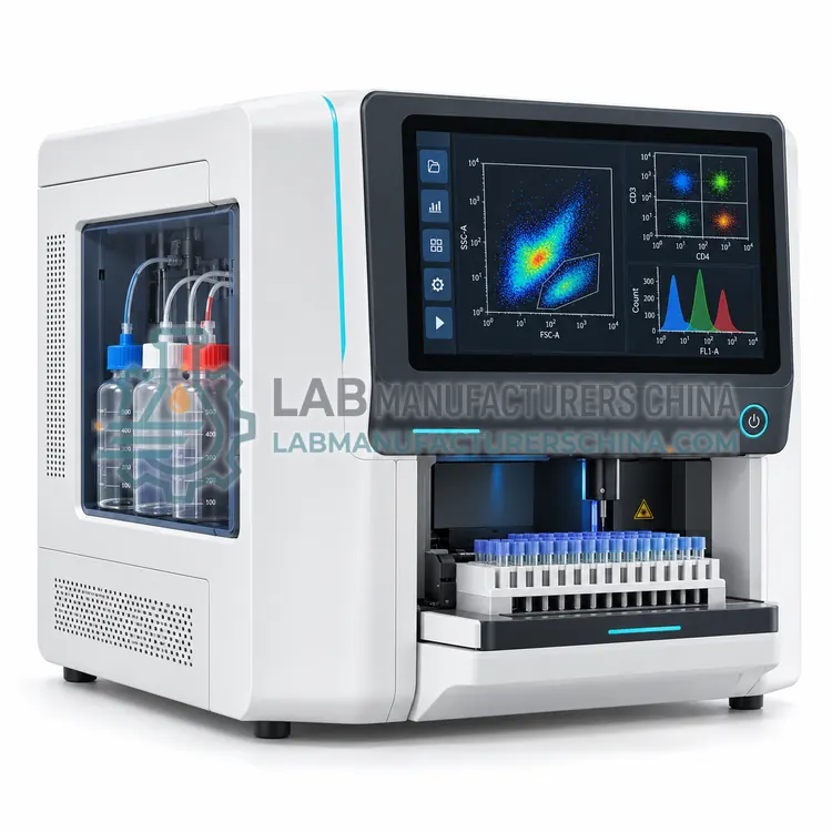
How do Flow Cytometers work?
A flow cytometer uses sheath fluid to hydrodynamically focus a sample into a narrow core stream so cells cross the laser interrogation point one at a time. Scattered light is collected at small angles (forward scatter, related to size) and at 90 degrees (side scatter, related to granularity). Fluorochromes are excited by lasers, commonly 405, 488, 561 and 640 nm, and emission is split by dichroic filters and bandpass filters to photomultiplier tubes or photodiodes. Conventional instruments assign one detector per fluorochrome and apply a compensation matrix, whereas spectral instruments capture the full emission spectrum and unmix it mathematically. Electronics digitize pulse height, area and width, and software records list-mode FCS files for gating and statistical analysis.
Which Flow Cytometers variants are available?
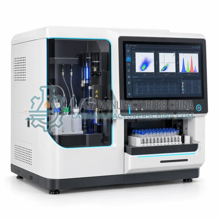
- Benchtop Flow Cytometer — A benchtop flow cytometer is a compact, filter-based analyzer with one to three lasers and roughly 4 to 10 fluorescence channels.
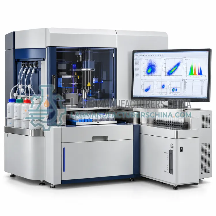
- High-Parameter Research Flow Cytometer — A high-parameter research flow cytometer is a modular analyzer with four to seven lasers and 18 to 30 or more fluorescence detectors.
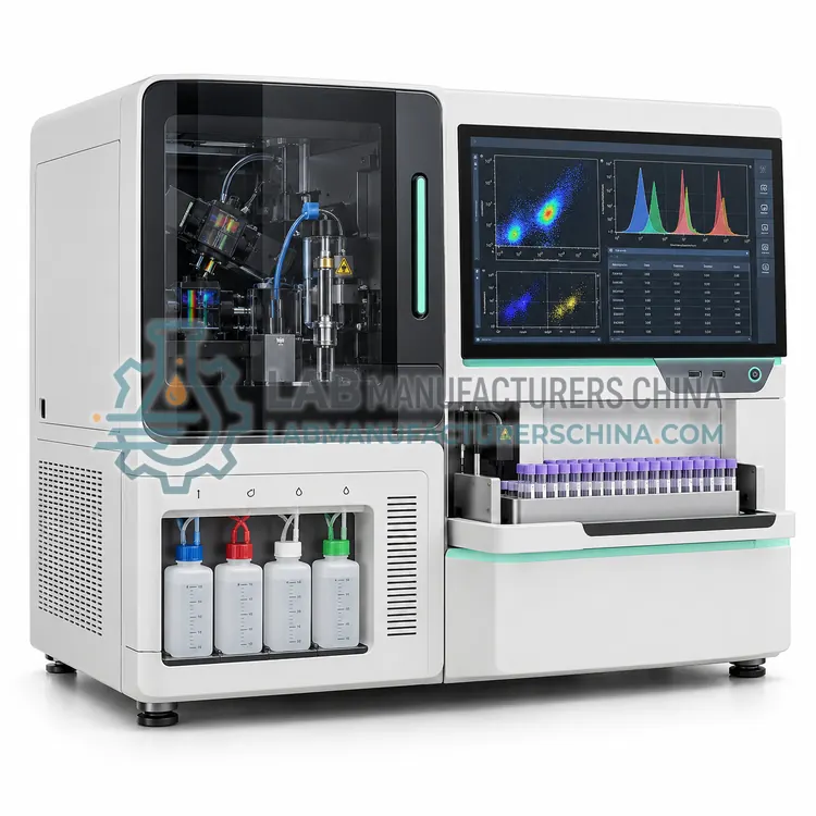
- Spectral Flow Cytometer — A spectral flow cytometer records the full fluorescence emission spectrum of every cell across an array of detectors and mathematically unmixes overlapping dyes.
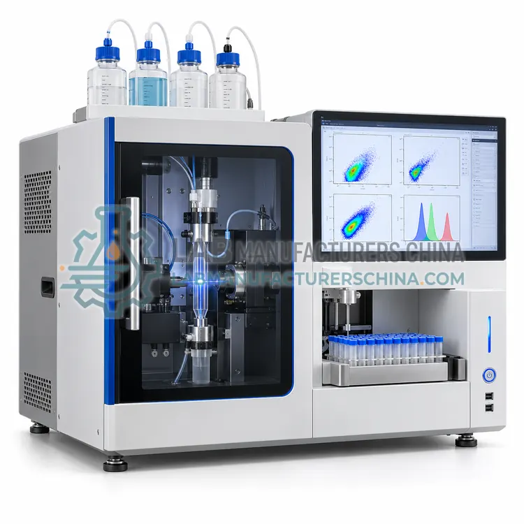
- Acoustic-Focusing Flow Cytometer — An acoustic-focusing flow cytometer uses ultrasonic standing waves to position cells in the center of the flow stream before laser interrogation.
- Compact Personal Flow Cytometer — A compact personal flow cytometer is a small, low-maintenance analyzer with one or two lasers and three to six fluorescence channels.
How do I choose the right variant?
Select a cytometer by panel complexity, sensitivity and workflow. Define the maximum number of simultaneous colors, then choose laser and detector count to match; conventional filter-based systems are simple for 4 to 12 colors, while spectral systems ease panel design beyond 20. Check resolution of dim populations, small particle detection and event rate, typically 10,000 to 100,000 events per second. Consider footprint, whether a compact personal analyzer or a full research platform is required, and whether an acoustic focusing or plate loader is needed for throughput. Verify that software provides compensation or unmixing, batch analysis and user access control. For clinical use, confirm that the supplier can support IVD documentation, calibration beads and a validated quality control routine.
Where are Flow Cytometers used?
Flow Cytometers are specified in immunophenotyping of lymphocyte subsets, CD34 stem cell enumeration, cell cycle and apoptosis analysis, leukemia and lymphoma diagnostics research, cell line and CAR-T characterization and bacterial and microbial counting.
Which standards and certifications apply?
Work with Flow Cytometers is commonly governed by CLSI H62, CLSI H43, ISO 20391-1, IEC 61010-1, IEC 61010-2-101, IEC 60825-1 and ISAC FCS 3.1. Instrument certifications such as CE marking are confirmed per configuration and never assumed.
What are the key considerations before purchase?
Install the cytometer on a vibration-free bench with stable power and ambient temperature between 15 and 30 degrees Celsius, and provide waste handling for potentially biohazardous effluent, typically with bleach treatment. Run daily quality control using fluorescent beads to verify laser alignment, sensitivity and coefficient of variation, and keep Levey-Jennings style records of detector voltages. Perform daily and weekly fluidics cleaning to avoid clogging, and replace sheath filters and tubing on schedule. Laser power and optical alignment should be checked by trained service staff at least annually. Laboratories working under ISO 15189 or GLP should enable audit trails, electronic signatures and validated software updates.