How do Clinical Microscopy work?
A compound microscope forms an enlarged image in two stages. The objective lens creates a real intermediate image of the specimen, and the eyepiece magnifies it further, giving total magnification equal to objective power times eyepiece power, commonly 40× to 1,000×. A condenser focuses light from an LED or halogen source onto the specimen using Köhler illumination for even contrast. Phase-contrast optics convert small refractive index differences in live cells into intensity differences using a phase ring. Dark-field blocks direct light so only scattered light enters the objective, making thin organisms bright on dark background. Fluorescence microscopes use an excitation filter, dichroic mirror and emission filter to show fluorophore-labeled targets. Digital systems add a camera or scanner that captures images and stitches fields into whole slide images.
Which Clinical Microscopy variants are available?
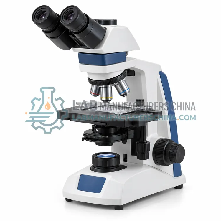
- Brightfield Clinical Binocular Microscope — A brightfield clinical binocular microscope illuminates stained specimens from below and is used for blood film differentials, Gram stains, acid-fast smears and tissue sections.
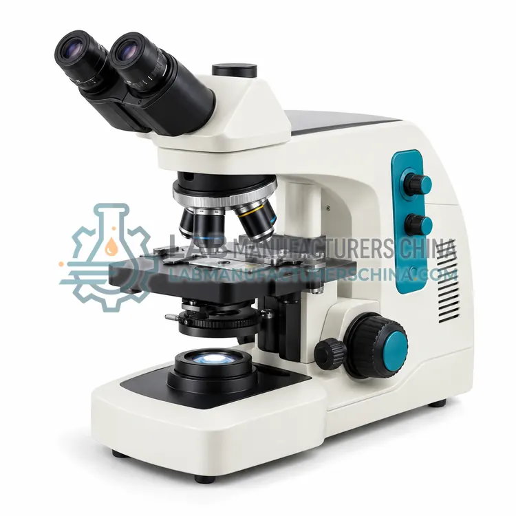
- Phase-Contrast Microscope for Urine Sediment and Semen — A phase-contrast microscope for urine sediment and semen uses phase rings to reveal unstained transparent cells and casts without staining.
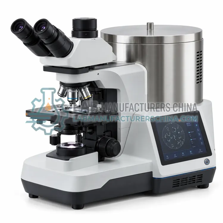
- LED Fluorescence Microscope for Clinical Diagnostics — An LED fluorescence microscope excites fluorescent dyes or labeled antibodies with LED light and images them through barrier filters.
- Dark-Field Microscope for Spirochete Detection — A dark-field microscope illuminates the specimen with oblique light so that only scattered light enters the objective, making thin unstained organisms appear bright on a dark background.
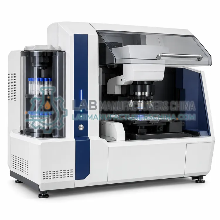
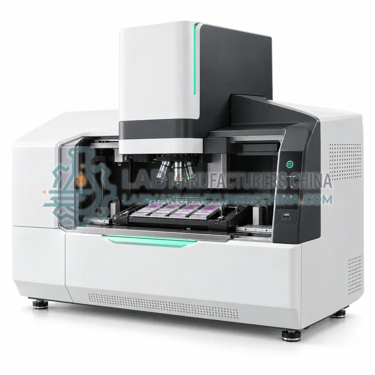
- Digital Whole Slide Scanner — A digital whole slide scanner images an entire glass slide at high resolution by scanning and stitching fields, creating a virtual slide that can be viewed, archived and shared.
How do I choose the right variant?
Choose the microscope by specimen type and contrast technique. Brightfield with plan achromat objectives suits stained blood films, Gram stains and histology. Phase contrast is preferred for unstained urine sediment, semen and live cell culture. Dark-field suits spirochetes such as Treponema pallidum, and fluorescence suits auramine acid-fast screening and immunofluorescence. Check objective quality, numerical aperture, field number, stage mechanics, illumination life and whether a camera port or digital system is needed for documentation and telepathology. Consider ergonomic features such as tilting binocular heads and low stage height for long screening sessions, and confirm objective parfocality and availability of spare lamps and service support.
Where are Clinical Microscopy used?
Clinical Microscopy are specified in blood film differential examination, urine sediment examination, microbiology smear examination, cytology and histopathology screening and semen analysis.
Which standards and certifications apply?
Work with Clinical Microscopy is commonly governed by ISO 8039, ISO 10110, IEC 61010-1, ISO 15189, ISO 19012-1 and ISO 19012-2. Instrument certifications such as CE marking are confirmed per configuration and never assumed.
What are the key considerations before purchase?
Place the microscope on a stable, vibration-free bench away from direct sunlight and dust, and use a cover when idle. Clean lenses only with lens tissue and approved solvent, and remove immersion oil after each use. Align Köhler illumination and verify parfocality at installation and after servicing. Calibrate eyepiece reticles or camera scale with a stage micrometer for measurements. Replace LED or halogen sources according to lifetime, and inspect objectives for scratches and fungus in humid climates. Staff should observe ergonomic posture and breaks. For digital systems, validate image quality and storage integrity in line with laboratory procedures under ISO 15189.