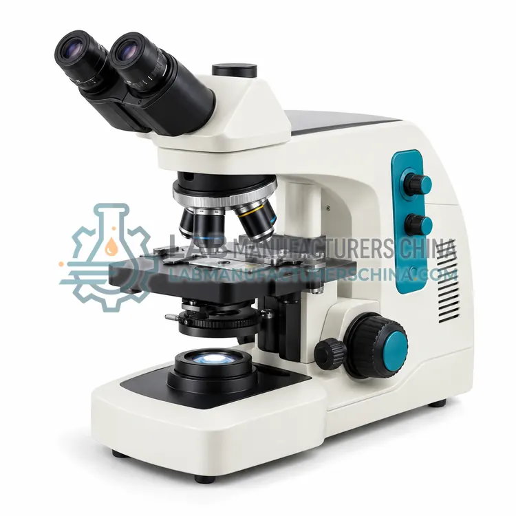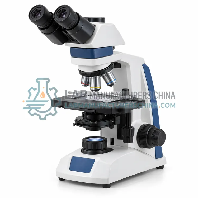
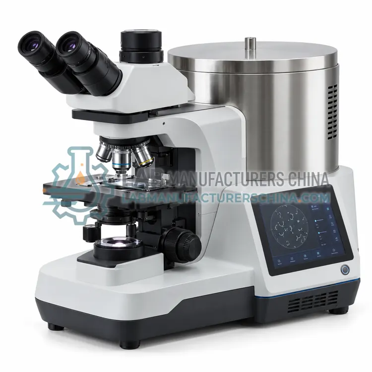
Dark-Field Microscope for Spirochete Detection
螺旋体检测暗视野显微镜
- SKU CLM-0004
- Clinical Microscopy
- CE (to be confirmed)
- ISO 13485 quality system (to be confirmed)
Price on request — FOB / CIF quoted per order
Prices depend on configuration, quantity and destination port.
Key specifications
| Observation method | Dark-field (and brightfield) |
|---|---|
| Condenser | Dry/oil dark-field, NA 1.20–1.43 |
| Objectives | 10×/40×/100× (iris for 100×) |
| Magnification | 100×–1,000× |
| Illumination | High-intensity LED, adjustable |
| Head | Binocular or trinocular |
Request a quote Download brochure · to be provided
By Lab Manufacturers China Technical TeamLast reviewed Standards: ISO 8039, ISO 10110, IEC 61010-1
What is the Dark-Field Microscope for Spirochete Detection?
A dark-field microscope illuminates the specimen with oblique light so that only scattered light enters the objective, making thin unstained organisms appear bright on a dark background. It is used for direct detection of motile Treponema pallidum in lesion exudate.
What is the Dark-Field Microscope for Spirochete Detection?
A dark-field microscope illuminates the specimen with oblique light so that only scattered light enters the objective, making thin unstained organisms appear bright on a dark background. It is used for direct detection of motile Treponema pallidum in lesion exudate.
Overview
A dark-field condenser contains a central stop that blocks direct light and sends a hollow cone of light at steep angles onto the specimen. Only light scattered or diffracted by structures enters the objective, so organisms as thin as 0.15 µm, below the resolution limit of ordinary brightfield, produce bright signals on a black field. This variant uses a dry or oil dark-field condenser with numerical aperture of 1.20 to 1.43, used with an objective whose numerical aperture is lower than the condenser, often 40× or 100× with iris diaphragm to reduce aperture. Immediate examination of fresh lesion material is required because motility is essential for identification. Strong LED illumination with intensity control and clean slides reduce background glare. Some microscopes combine dark-field with brightfield and phase contrast.
How does it work?
A compound microscope forms an enlarged image in two stages. The objective lens creates a real intermediate image of the specimen, and the eyepiece magnifies it further, giving total magnification equal to objective power times eyepiece power, commonly 40× to 1,000×. A condenser focuses light from an LED or halogen source onto the specimen using Köhler illumination for even contrast. Phase-contrast optics convert small refractive index differences in live cells into intensity differences using a phase ring. Dark-field blocks direct light so only scattered light enters the objective, making thin organisms bright on dark background. Fluorescence microscopes use an excitation filter, dichroic mirror and emission filter to show fluorophore-labeled targets. Digital systems add a camera or scanner that captures images and stitches fields into whole slide images.
What are the key features?
- Dark-field condenser with NA 1.20–1.43 for oblique illumination
- Detects organisms thinner than 0.2 µm on a black background
- Objectives with iris diaphragm to lower numerical aperture
- Strong LED illumination with intensity control
- Suitable for live motility observation of fresh specimens
- Optional combination with brightfield and phase contrast
What are the technical specifications?
A typical dark-field microscope for spirochete detection configuration offers observation method of Dark-field (and brightfield), condenser of Dry/oil dark-field, NA 1.20–1.43, objectives of 10×/40×/100× (iris for 100×), magnification of 100×–1,000×, illumination of High-intensity LED, adjustable and head of Binocular or trinocular. Values are indicative of the product class; the exact figures for each configuration are confirmed on the quotation and the calibration documents supplied with the instrument.
Which applications is it used for?
The Dark-Field Microscope for Spirochete Detection is used in treponema pallidum detection in lesion exudate, leptospira examination, live motile microorganism observation and teaching and research. Across the wider Clinical Microscopy class, laboratories also deploy it for blood film differential examination, urine sediment examination and microbiology smear examination.
Which standards and certifications apply?
Relevant standards and methods include ISO 8039, ISO 10110, IEC 61010-1, ISO 15189, ISO 19012-1 and ISO 19012-2. Certification candidates for this model are CE and ISO 13485 quality system; each is to be confirmed for the specific configuration before shipment, and certificates are supplied only where they have been issued.
Place the microscope on a stable, vibration-free bench away from direct sunlight and dust, and use a cover when idle. Clean lenses only with lens tissue and approved solvent, and remove immersion oil after each use. Align Köhler illumination and verify parfocality at installation and after servicing. Calibrate eyepiece reticles or camera scale with a stage micrometer for measurements. Replace LED or halogen sources according to lifetime, and inspect objectives for scratches and fungus in humid climates. Staff should observe ergonomic posture and breaks. For digital systems, validate image quality and storage integrity in line with laboratory procedures under ISO 15189.
How do I select the right Dark-Field Microscope for Spirochete Detection?
Choose this variant where direct detection of motile spirochetes is required, such as in sexually transmitted infection clinics. In the RFQ specify dark-field condenser type and numerical aperture, iris objective, illumination intensity, oil or dry condenser and camera port. Serology is still needed to confirm diagnosis when direct examination is negative.
How does it compare with other Clinical Microscopy variants?
Choose the microscope by specimen type and contrast technique. Brightfield with plan achromat objectives suits stained blood films, Gram stains and histology. Phase contrast is preferred for unstained urine sediment, semen and live cell culture. Dark-field suits spirochetes such as Treponema pallidum, and fluorescence suits auramine acid-fast screening and immunofluorescence. Check objective quality, numerical aperture, field number, stage mechanics, illumination life and whether a camera port or digital system is needed for documentation and telepathology. Consider ergonomic features such as tilting binocular heads and low stage height for long screening sessions, and confirm objective parfocality and availability of spare lamps and service support.
Full specifications
| Observation method | Dark-field (and brightfield) |
|---|---|
| Condenser | Dry/oil dark-field, NA 1.20–1.43 |
| Objectives | 10×/40×/100× (iris for 100×) |
| Magnification | 100×–1,000× |
| Illumination | High-intensity LED, adjustable |
| Head | Binocular or trinocular |
| Eyepieces | WF10×/20 mm |
| Stage travel | 75 × 50 mm |
| Focus | Coaxial coarse/fine |
| Camera port | Optional C-mount |
| Power supply | 100–240 V AC, 50/60 Hz |
Indicative values for this product class. Final specifications are confirmed on the quotation and in the calibration documents supplied with the instrument.
Applications
- Treponema pallidum detection in lesion exudate
- Leptospira examination
- Live motile microorganism observation
- Teaching and research
Key features
- Dark-field condenser with NA 1.20–1.43 for oblique illumination
- Detects organisms thinner than 0.2 µm on a black background
- Objectives with iris diaphragm to lower numerical aperture
- Strong LED illumination with intensity control
- Suitable for live motility observation of fresh specimens
- Optional combination with brightfield and phase contrast
Frequently asked questions about Dark-Field Microscope for Spirochete Detection
Dark-field microscopy shows thin, motile organisms such as Treponema pallidum and Leptospira, which are invisible in ordinary brightfield unstained wet mounts. Identification depends on characteristic corkscrew morphology and motility, so examination must be performed on fresh material.
Dark-field shows specimens as bright objects on a black background using scattered light from oblique illumination, while phase contrast shows internal detail as shades of gray using phase rings. Dark-field excels for thin organisms, and phase contrast for cell structure.
Slides must be clean, thin (about 1.0 mm) and free of scratches and dust, since any particle scatters light. Coverslips should be thin and of consistent thickness, and immersion oil, if used on the condenser, must be free of bubbles.
It is used to examine specimens under magnification for diagnosis, including blood cell morphology, urine sediment, parasites, bacteria in stained smears, cytology and tissue sections. Different microscope types provide brightfield, phase-contrast, dark-field or fluorescence contrast for different specimens.
Standard objectives are 4×, 10×, 40× and 100× oil immersion, paired with 10× eyepieces to give 40× to 1,000× total magnification. Low power locates fields, 40× is used for urine sediment and cytology, and 100× oil is used for blood films and bacterial morphology.
Köhler illumination is a method of adjusting the field and condenser apertures so the specimen is evenly lit and glare is minimized. It gives maximum resolution and contrast from the objective, and should be set after installing or moving the microscope and when changing objectives.
Related models