Phase-Contrast Microscope for Urine Sediment and Semen
尿沉渣和精液检查相差显微镜
- SKU CLM-0002
- Clinical Microscopy
- CE (to be confirmed)
- ISO 13485 quality system (to be confirmed)
Price on request — FOB / CIF quoted per order
Prices depend on configuration, quantity and destination port.
Key specifications
| Observation method | Phase contrast and brightfield |
|---|---|
| Magnification | 100×–400× |
| Phase objectives | 10×/20×/40× (PH1/PH2) |
| Condenser | Turret with phase annuli, NA 0.9 |
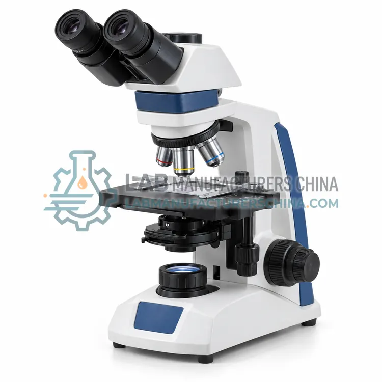
| Head | Binocular or trinocular |
| Eyepieces | WF10×/20 mm |
Request a quote Download brochure · to be provided
By Lab Manufacturers China Technical TeamLast reviewed Standards: ISO 8039, ISO 10110, IEC 61010-1
What is the Phase-Contrast Microscope for Urine Sediment and Semen?
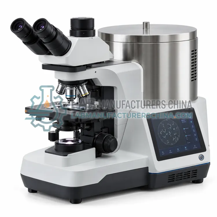
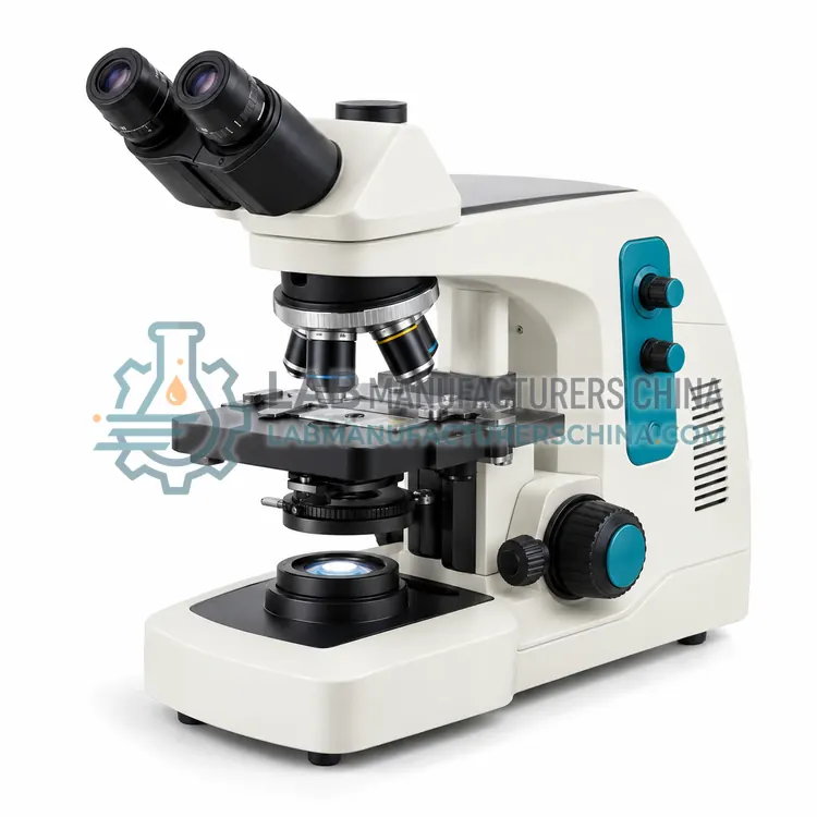
A phase-contrast microscope for urine sediment and semen uses phase rings to reveal unstained transparent cells and casts without staining. It is used in urinalysis, andrology and cell culture laboratories to see low-contrast structures such as hyaline casts and sperm motility.
What is the Phase-Contrast Microscope for Urine Sediment and Semen?
A phase-contrast microscope for urine sediment and semen uses phase rings to reveal unstained transparent cells and casts without staining. It is used in urinalysis, andrology and cell culture laboratories to see low-contrast structures such as hyaline casts and sperm motility.
Overview
Phase contrast converts phase shifts created by differences in refractive index into brightness differences. An annular diaphragm in the condenser produces a hollow cone of light, and a matching phase ring in the objective shifts the phase of the direct light by a quarter wavelength and dims it, so that diffracted light from the specimen interferes and creates contrast. This variant typically carries phase objectives at 10×, 20× and 40× with matching condenser annuli in a turret, plus a brightfield position. It resolves hyaline casts, renal tubular cells, yeast, and unstained sperm motility in wet mounts. A heated stage at 37 °C is available for semen analysis. LED illumination with green filter improves contrast, and a camera port lets the laboratory document findings and compute sperm concentration using a counting chamber.
How does it work?
A compound microscope forms an enlarged image in two stages. The objective lens creates a real intermediate image of the specimen, and the eyepiece magnifies it further, giving total magnification equal to objective power times eyepiece power, commonly 40× to 1,000×. A condenser focuses light from an LED or halogen source onto the specimen using Köhler illumination for even contrast. Phase-contrast optics convert small refractive index differences in live cells into intensity differences using a phase ring. Dark-field blocks direct light so only scattered light enters the objective, making thin organisms bright on dark background. Fluorescence microscopes use an excitation filter, dichroic mirror and emission filter to show fluorophore-labeled targets. Digital systems add a camera or scanner that captures images and stitches fields into whole slide images.
What are the key features?
- Phase-contrast objectives 10×, 20× and 40× with turret annuli
- Brightfield and phase modes in one condenser turret
- Optional 37 °C heated stage for semen analysis
- Green filter and LED illumination to enhance phase contrast
- Camera port for documentation and sperm counting
- Long working distance objectives for wet mounts and chambers
What are the technical specifications?
A typical phase-contrast microscope for urine sediment and semen configuration offers observation method of Phase contrast and brightfield, magnification of 100×–400×, phase objectives of 10×/20×/40× (PH1/PH2), condenser of Turret with phase annuli, NA 0.9, head of Binocular or trinocular and eyepieces of WF10×/20 mm. Values are indicative of the product class; the exact figures for each configuration are confirmed on the quotation and the calibration documents supplied with the instrument.
Which applications is it used for?
The Phase-Contrast Microscope for Urine Sediment and Semen is used in urine sediment examination of unstained specimens, semen motility and concentration, live cell culture monitoring and body fluid cell detection. Across the wider Clinical Microscopy class, laboratories also deploy it for blood film differential examination, urine sediment examination and microbiology smear examination.
Which standards and certifications apply?
Relevant standards and methods include ISO 8039, ISO 10110, IEC 61010-1, ISO 15189, ISO 19012-1 and ISO 19012-2. Certification candidates for this model are CE and ISO 13485 quality system; each is to be confirmed for the specific configuration before shipment, and certificates are supplied only where they have been issued.
Place the microscope on a stable, vibration-free bench away from direct sunlight and dust, and use a cover when idle. Clean lenses only with lens tissue and approved solvent, and remove immersion oil after each use. Align Köhler illumination and verify parfocality at installation and after servicing. Calibrate eyepiece reticles or camera scale with a stage micrometer for measurements. Replace LED or halogen sources according to lifetime, and inspect objectives for scratches and fungus in humid climates. Staff should observe ergonomic posture and breaks. For digital systems, validate image quality and storage integrity in line with laboratory procedures under ISO 15189.
How do I select the right Phase-Contrast Microscope for Urine Sediment and Semen?
Choose this variant when specimens are unstained or wet-mounted, as in urine sediment, semen and parasite screening. In the RFQ specify phase objective set and ring type, condenser turret positions, heated stage, filters, camera port and compatibility with counting chambers. Brightfield-only scopes cannot reveal low-contrast structures.
How does it compare with other Clinical Microscopy variants?
Choose the microscope by specimen type and contrast technique. Brightfield with plan achromat objectives suits stained blood films, Gram stains and histology. Phase contrast is preferred for unstained urine sediment, semen and live cell culture. Dark-field suits spirochetes such as Treponema pallidum, and fluorescence suits auramine acid-fast screening and immunofluorescence. Check objective quality, numerical aperture, field number, stage mechanics, illumination life and whether a camera port or digital system is needed for documentation and telepathology. Consider ergonomic features such as tilting binocular heads and low stage height for long screening sessions, and confirm objective parfocality and availability of spare lamps and service support.
Full specifications
| Observation method | Phase contrast and brightfield |
|---|---|
| Magnification | 100×–400× |
| Phase objectives | 10×/20×/40× (PH1/PH2) |
| Condenser | Turret with phase annuli, NA 0.9 |
| Head | Binocular or trinocular |
| Eyepieces | WF10×/20 mm |
| Illumination | LED with green filter |
| Heated stage | Optional, ambient to 40 °C ±0.5 °C |
| Stage travel | 75 × 50 mm |
| Focus | Coaxial coarse and fine |
| Power supply | 100–240 V AC, 50/60 Hz |
Indicative values for this product class. Final specifications are confirmed on the quotation and in the calibration documents supplied with the instrument.
Applications
- Urine sediment examination of unstained specimens
- Semen motility and concentration
- Live cell culture monitoring
- Body fluid cell detection
Key features
- Phase-contrast objectives 10×, 20× and 40× with turret annuli
- Brightfield and phase modes in one condenser turret
- Optional 37 °C heated stage for semen analysis
- Green filter and LED illumination to enhance phase contrast
- Camera port for documentation and sperm counting
- Long working distance objectives for wet mounts and chambers
Frequently asked questions about Phase-Contrast Microscope for Urine Sediment and Semen
It reveals transparent, unstained structures such as hyaline casts, epithelial cells, yeast and motile sperm by converting small refractive-index differences into contrast. Brightfield shows these poorly unless they are stained, which can distort or kill live specimens.
Phase objectives have a built-in phase ring, labeled PH1, PH2 or PH3 according to ring size, and must be matched to the corresponding condenser annulus. Common powers are 10×, 20× and 40×, and a brightfield objective may be used for 100× oil examination.
It is designed for unstained specimens and gives less value on stained slides, where brightfield is better. A phase microscope with a brightfield position lets the same instrument handle both, which is why most clinical phase models include a switchable condenser.
It is used to examine specimens under magnification for diagnosis, including blood cell morphology, urine sediment, parasites, bacteria in stained smears, cytology and tissue sections. Different microscope types provide brightfield, phase-contrast, dark-field or fluorescence contrast for different specimens.
Standard objectives are 4×, 10×, 40× and 100× oil immersion, paired with 10× eyepieces to give 40× to 1,000× total magnification. Low power locates fields, 40× is used for urine sediment and cytology, and 100× oil is used for blood films and bacterial morphology.
Köhler illumination is a method of adjusting the field and condenser apertures so the specimen is evenly lit and glare is minimized. It gives maximum resolution and contrast from the objective, and should be set after installing or moving the microscope and when changing objectives.
Related models