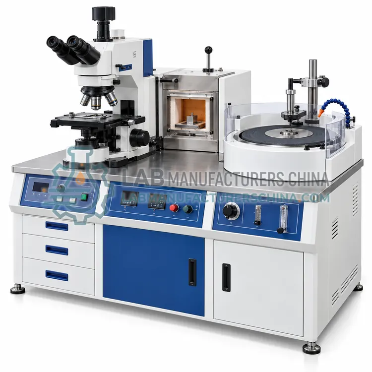
How do Microscopy work?
A metallurgical microscope illuminates the specimen from the same side as the objective, since metal is opaque, and the polished and etched surface reflects light back through the optical system. Contrast comes from differences in reflectivity between phases, from surface relief created by etching, and from scattering by inclusions and cracks. Objective numerical aperture determines resolution, and magnification should be chosen so that the features of interest occupy a reasonable fraction of the field, because empty magnification adds no detail. Illumination modes extend the range: brightfield for general structure, darkfield for inclusions and cracks, polarized light for anisotropic phases and coatings, and differential interference contrast for relief and small height differences. Stereo microscopes use separate light paths for each eye to give depth perception, which makes them suitable for fracture surfaces and weld profiles. Digital cameras and measuring software add quantitative output, such as grain size, coating thickness and defect dimensions.
Which Microscopy variants are available?
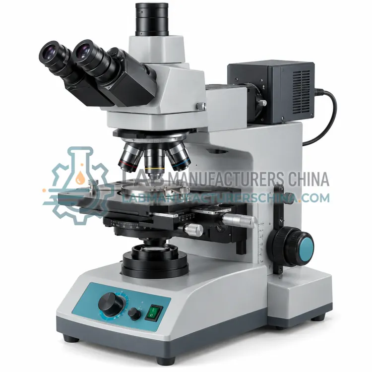
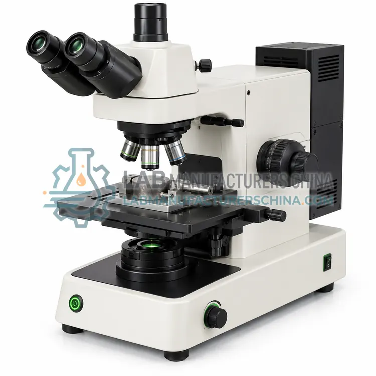
- Upright Reflected-Light Metallurgical Microscope — An upright reflected-light metallurgical microscope views polished and etched specimens from above with a full set of objectives and several illumination modes.
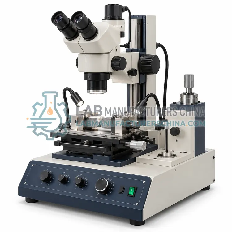
- Stereo Zoom Microscope for Fracture and Weld Examination — A stereo zoom microscope provides two slightly different views to give depth perception, making it suited to fracture surfaces, weld profiles and corrosion damage at low to medium magnification.
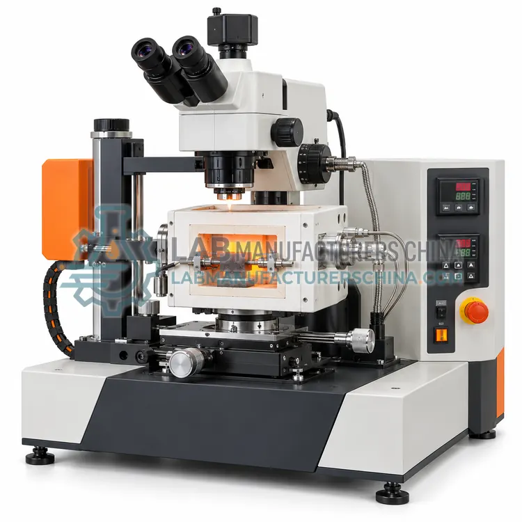
- Digital Vision Measuring Microscope — A digital vision measuring microscope combines optics with a calibrated camera and software to measure dimensions on small parts, tool edges, holes and coating cross-sections.
- Portable Field Metallurgical Microscope — A portable field metallurgical microscope examines metal surfaces in situ using a magnetic or clamp mount, often on replicas or prepared spots.
- Polarizing Light Microscope for Ore and Inclusion Study — A polarizing light microscope uses crossed polarizers to identify transparent minerals and anisotropic phases by their optical behaviour, and to characterise non-metallic inclusions.
How do I choose the right variant?
Choose the microscope type from what must be seen. Upright reflected-light instruments with a full objective set and multiple illumination modes handle polished sections, grain size and inclusion rating. Inverted stages are better for large or unmounted specimens. Stereo microscopes are the right tool for fracture surfaces, weld profiles, corrosion damage and assembly inspection, where depth and surface shape matter more than high magnification. Digital measuring microscopes suit dimensional work on small features, such as tool edges, holes, threads and coating cross-sections, where accuracy claims need to be supported by calibration. Portable microscopes serve on-site replica examination of installed plant. In every case, match the magnification range, numerical aperture and illumination modes to the features of interest, and budget for a stage micrometer, calibration slides and a camera whose resolution genuinely exceeds the detail the optics can deliver.
Where are Microscopy used?
Microscopy are specified in grain size and inclusion rating of steels, weld and heat-affected zone examination, fracture surface and failure investigation, coating and surface defect measurement and ore, slag and inclusion mineralogy.
Which standards and certifications apply?
Work with Microscopy is commonly governed by ASTM E3, ASTM E407, ASTM E112, ASTM E45, ISO 643, ASTM E1951 and IEC 61010-1. Instrument certifications such as CE marking are confirmed per configuration and never assumed.
What are the key considerations before purchase?
Clean lenses only with the recommended method, since immersion oils and abrasives damage coatings, and cover the instrument when not in use. Verify magnification and length calibration with a stage micrometer at each objective and after any change of camera or software, and keep a record with the results. Set illumination to a reproducible level, because contrast in reflected light depends strongly on lamp intensity and aperture settings, and note the objective and illumination mode in every report. Keep specimens clean and dry, store them in labelled containers, and use replicas rather than marking the component when examining installed parts. Service the illuminator, stage and focusing mechanism periodically, and check the camera sensor for dust that would appear as artefacts in images used for measurement, since a mistaken artefact can change a reported coating thickness.