Metallography reveals the internal structure of a metal by preparing a flat, scratch-free surface and then accentuating features chemically or optically. A cut-off wheel sections the specimen under coolant so heat does not alter the structure, and hot or cold mounting encapsulates it in resin for handling. Grinding with silicon carbide papers removes the cut damage, and polishing with diamond or alumina suspensions produces a mirror-like surface. Etching with a reagent such as nital attacks grain boundaries and specific phases preferentially, so reflected light shows grain structure, phase contrast and defects. Examination uses inverted or upright reflected-light microscopes with brightfield, darkfield, polarized and differential interference contrast modes, and images are captured digitally. Image analysis then measures grain size by intercept or planimetric methods, phase fractions, nodularity, inclusion ratings and coating thickness, producing numbers that can be compared against acceptance limits.
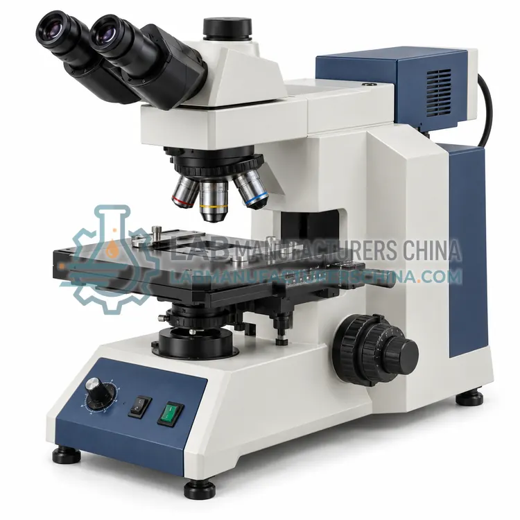
- Inverted Metallographic Microscope — An inverted metallographic microscope observes the polished surface of a specimen from below, so large or heavy unmounted samples rest on the stage.
- Metallographic Image Analysis System — A metallographic image analysis system captures microscope images and applies calibrated measurement routines to quantify grain size, phase fractions, nodularity, inclusion ratings and coating thickness.
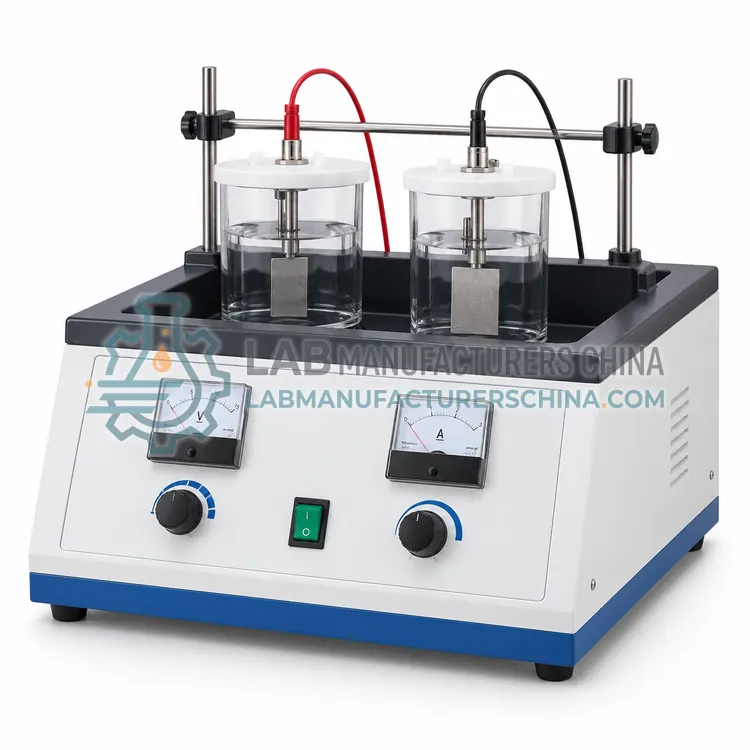
- Electrolytic Polishing and Etching Unit — An electrolytic polishing and etching unit passes a controlled current through a specimen immersed in an electrolyte, dissolving the surface uniformly to polish it or selectively to reveal microstructure.
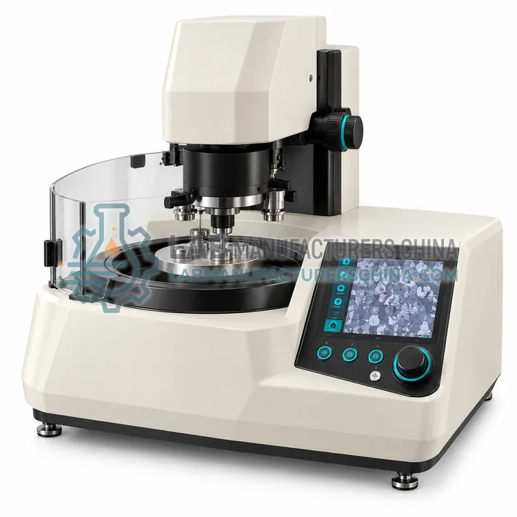
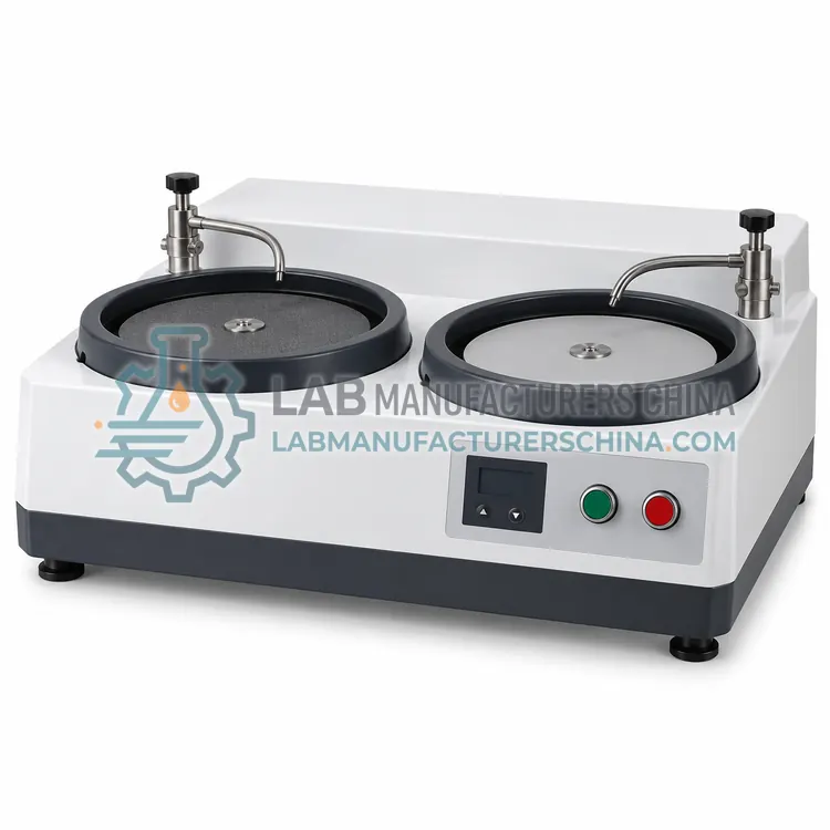
- Metallographic Specimen Grinding and Polishing Machine — A metallographic grinding and polishing machine rotates abrasive papers and cloths while holding specimens under controlled force, removing cutting damage and producing a scratch-free surface.
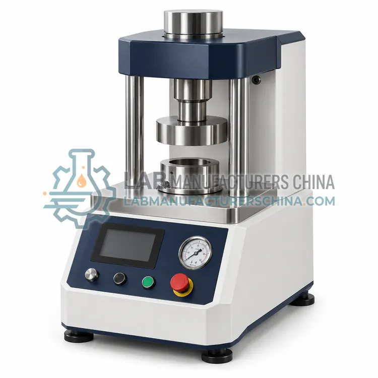
- Hot Mounting Press for Metallographic Specimens — A hot mounting press encapsulates a cut metallographic specimen in thermosetting resin under heat and pressure, producing a uniform cylindrical mount that protects edges and suits automatic polishing.
How do I choose the right variant?
Choose preparation equipment by specimen type and daily volume. Hardened steel and large sections need a sturdy cut-off saw with adequate cooling, while small or porous parts need vacuum impregnation or cold mounting. A semi-automatic grinding and polishing head with individual force control gives reproducible results for several specimens at once and is worth the cost when many samples are prepared daily. For examination, decide whether an upright microscope is enough or whether an inverted stage is needed for large, unmounted samples, and check the magnification range, illumination modes and camera resolution against the features you measure. Image analysis software should support the specific measurements you report, such as ASTM E112 grain size, ASTM E45 inclusion ratings or coating thickness, and it should allow the measurement rules and thresholds to be recorded so results can be audited and reproduced by another operator.
Metallographic Equipment are specified in microstructure examination in steel and cast iron, grain size and phase fraction measurement, inclusion and cleanliness rating, coating and case depth measurement and weld and heat-affected zone examination.
Which standards and certifications apply?
Work with Metallographic Equipment is commonly governed by ASTM E3, ASTM E407, ASTM E112, ASTM E45, ASTM E562, ISO 643 and IEC 61010-1. Instrument certifications such as CE marking are confirmed per configuration and never assumed.
What are the key considerations before purchase?
Consumables dominate metallographic quality, so keep an adequate stock of cut-off wheels, resin, papers, cloths and diamond suspensions, and follow the supplier's recommended sequence. Clean specimens thoroughly between steps with water and alcohol and dry them before etching, since contamination produces artefacts. Prepare a batch of control specimens of known structure to check that the procedure still reveals the true structure, and date all etchants, replacing them when they weaken. Verify microscope calibration with a stage micrometer each time magnification is changed, and confirm that the software's length scale matches the objective actually used. Handle etchants and solvents with gloves, eye protection and fume extraction, and dispose of spent solutions through an authorised route, since many contain strong acids or organic solvents.