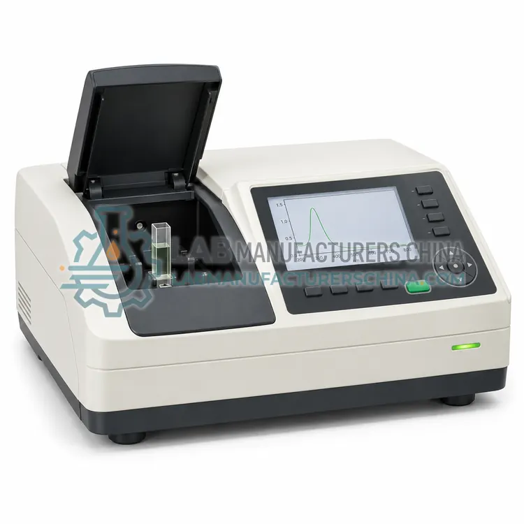
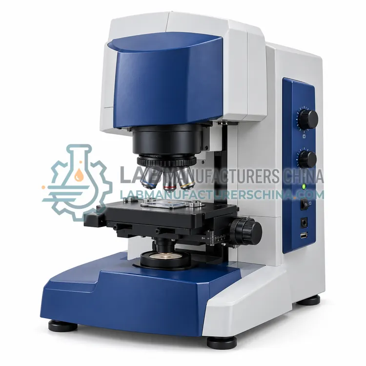
FTIR Microscope for Pharmaceutical Particle Analysis
制药颗粒分析红外显微镜
- SKU FTR-0005
- FTIR
- CE (to be confirmed)
- IEC 61010-1 compliance (to be confirmed)
- ISO 13485 manufacturing (to be confirmed)
Price on request — FOB / CIF quoted per order
Prices depend on configuration, quantity and destination port.
Key specifications
| Spatial resolution | about 10–50 µm |
|---|---|
| Spectral range | 4000–650 cm-1 |
| Resolution | 4–8 cm-1 |
| Sampling modes | Transmission and reflectance |
| Sample cells | Compression or diamond cell |
| Magnification | about 15× to 30× |
Request a quote Download brochure · to be provided
By Lab Manufacturers China Technical TeamLast reviewed Standards: USP <854>, USP <197>, USP <197M>
What is the FTIR Microscope for Pharmaceutical Particle Analysis?
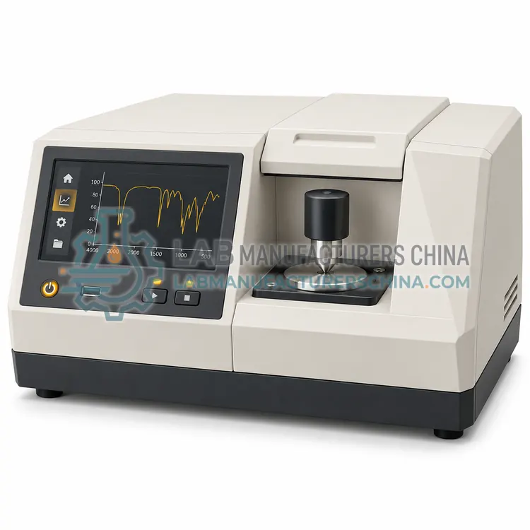
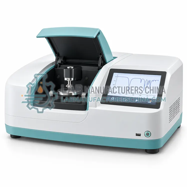
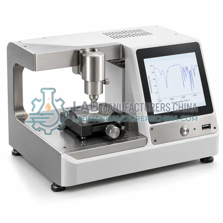
An FTIR microscope couples an infrared spectrometer to an optical microscope so individual particles and contaminants can be identified. It is chosen for investigating foreign matter, defects and small inclusions found in batches and finished products.
What is the FTIR Microscope for Pharmaceutical Particle Analysis?
An FTIR microscope couples an infrared spectrometer to an optical microscope so individual particles and contaminants can be identified. It is chosen for investigating foreign matter, defects and small inclusions found in batches and finished products.
Overview
An FTIR microscope focuses infrared light through microscope optics onto a small area, often tens of micrometres across, so a single particle can be measured and identified. The analyst first locates the particle visually, then collects a spectrum, and compares it with a library of polymers, minerals, excipients and actives. Sampling may be transmission through a compression cell or a diamond cell, or reflectance, and some systems add an imaging detector that maps chemical composition across a sample area. Applications include identifying foreign particles in a batch, characterising inclusions in tablets, studying contaminant fibres and investigating packaging interactions. Because particles are small and often irregular, sample preparation and the ability to reposition the aperture precisely are critical.
How does it work?
An FTIR spectrometer shines broadband infrared light through an interferometer, usually a Michelson type with a moving mirror, and then through or onto the sample. The detector records an interferogram, which a Fourier transform converts into an absorption spectrum from about 4000 to 400 cm-1. Molecular bonds absorb at characteristic frequencies, so the spectrum acts as a fingerprint of the material. Sampling accessories determine how the sample is presented: transmission through a potassium bromide pellet or a liquid cell, attenuated total reflectance for solids and pastes, diffuse reflectance for powders, or a microscope for small particles. Resolution, signal-to-noise ratio, wavenumber accuracy and the condition of the sampling accessory determine whether a spectrum matches the reference.
What are the key features?
- Infrared beam focused onto a small area
- Optical viewing to locate the particle
- Transmission or reflectance sampling modes
- Library comparison for polymers and minerals
- Optional chemical imaging detector
- Precise aperture positioning on small samples
What are the technical specifications?
A typical ftir microscope for pharmaceutical particle analysis configuration offers spatial resolution of about 10–50 µm, spectral range of 4000–650 cm-1, resolution of 4–8 cm-1, sampling modes of Transmission and reflectance, sample cells of Compression or diamond cell and magnification of about 15× to 30×. Values are indicative of the product class; the exact figures for each configuration are confirmed on the quotation and the calibration documents supplied with the instrument.
Which applications is it used for?
The FTIR Microscope for Pharmaceutical Particle Analysis is used in foreign particle identification, tablet inclusion analysis, contaminant fibre analysis and packaging interaction studies. Across the wider FTIR class, laboratories also deploy it for raw material identification at goods-in, excipient identity confirmation and polymorph and crystal form screening.
Which standards and certifications apply?
Relevant standards and methods include USP <854>, USP <197>, USP <197M>, EP 2.2.24, ICH Q2(R2) and IEC 61010-1. Certification candidates for this model are CE, IEC 61010-1 compliance and ISO 13485 manufacturing; each is to be confirmed for the specific configuration before shipment, and certificates are supplied only where they have been issued.
Keep the instrument in a room with stable temperature and low humidity, and keep desiccant fresh, because water vapour and carbon dioxide produce sharp absorption bands that can obscure the fingerprint region. Align the interferometer and check wavenumber accuracy with a polystyrene film or other standard at defined intervals, and record the results. Clean ATR crystals carefully, since scratches and residues change the spectrum, and use a background scan at the same settings as the sample. Handle potassium bromide with a dry atmosphere because it is hygroscopic. Maintain the library and re-verify performance after any accessory or detector change, and retain raw spectra for inspection.
How do I select the right FTIR Microscope for Pharmaceutical Particle Analysis?
Specify an FTIR microscope when foreign matter or small inclusions must be identified and visual inspection alone is insufficient. Confirm spatial resolution, sampling modes, sample cell options, library coverage and imaging capability. Consider whether chemical mapping is needed for heterogeneous samples.
How does it compare with other FTIR variants?
Choose an FTIR system from the sample form and the test required. For raw material identity, an ATR accessory with a diamond or zinc selenide crystal is convenient because powders and pastes can be measured with minimal preparation, and a benchtop unit with a validated spectral library suits goods-in testing. For polymorph screening and structural confirmation, higher resolution and careful control of sample preparation are needed, and comparisons should use the same accessory and conditions as the reference. Portable or handheld FTIR instruments allow identification through packaging or at the point of sampling. FTIR microscopy is used for particle and contaminant identification. Compare spectral range, resolution, signal-to-noise ratio, accessory options and library software with audit trails.
Full specifications
| Spatial resolution | about 10–50 µm |
|---|---|
| Spectral range | 4000–650 cm-1 |
| Resolution | 4–8 cm-1 |
| Sampling modes | Transmission and reflectance |
| Sample cells | Compression or diamond cell |
| Magnification | about 15× to 30× |
| Imaging option | Chemical mapping detector |
| Power supply | 220 V, 50 Hz, about 0.4 kW |
Indicative values for this product class. Final specifications are confirmed on the quotation and in the calibration documents supplied with the instrument.
Applications
- Foreign particle identification
- Tablet inclusion analysis
- Contaminant fibre analysis
- Packaging interaction studies
Key features
- Infrared beam focused onto a small area
- Optical viewing to locate the particle
- Transmission or reflectance sampling modes
- Library comparison for polymers and minerals
- Optional chemical imaging detector
- Precise aperture positioning on small samples
Frequently asked questions about FTIR Microscope for Pharmaceutical Particle Analysis
Typically particles from about 10 to 50 micrometres can be measured, depending on the aperture, detector and how strongly the material absorbs. Smaller particles may give noisy spectra, and a diamond compression cell can improve contact and signal.
Particles are transferred to a suitable window or compression cell under a stereomicroscope, flattened where necessary to give a usable path length, and positioned in the infrared beam. Contamination during transfer must be avoided, since the particle is the sample.
It maps the distribution of different materials across a sample by collecting spectra at many points, which helps characterise layered structures, inclusions inside a tablet or mixtures of particles that cannot be separated for individual measurement.
The infrared spectrum of the sample is compared with a reference spectrum, a pharmacopoeial reference standard or a library entry. A match across the characteristic absorption bands confirms the molecular identity of the material under the defined sampling conditions.
ATR uses a crystal of high refractive index that the sample contacts. Infrared light undergoes total internal reflection inside the crystal and the evanescent wave probes a thin surface layer, so solids, pastes and liquids can be measured with little or no preparation.
Water and carbon dioxide absorb strongly in the infrared and create sharp rotational bands that can overlap the analyte spectrum, especially in the fingerprint region. Stable humidity, fresh desiccant and an appropriate background scan reduce this interference.
Related models