Benchtop Flow Cytometer
台式流式细胞仪
- SKU CEA-0004
- Cell Analysis
- CE (to be confirmed)
- ISO 9001 manufacturing (to be confirmed)
- IEC 60825-1 laser product classification (to be confirmed)
Price on request — FOB / CIF quoted per order
Prices depend on configuration, quantity and destination port.
Key specifications
| Lasers | 488 nm, optional 405 and 640 nm |
|---|---|
| Fluorescence channels | 2–8 colors |
| Scatter detection | FSC, SSC |
| Acquisition rate | up to 10,000–35,000 events/s |
| Sample flow rate | 10–120 μL/min |
| Fluorescence sensitivity | FITC below 500 MESF (typical) |
Request a quote Download brochure · to be provided
By Lab Manufacturers China Technical TeamLast reviewed Standards: IEC 61010-1, IEC 61010-2-101, IEC 61326-2-6
What is the Benchtop Flow Cytometer?
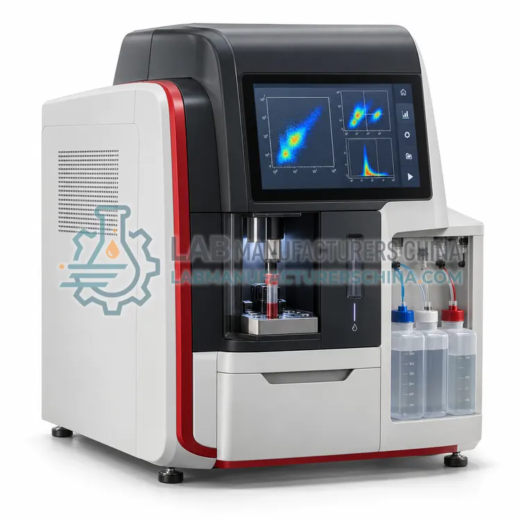
A benchtop flow cytometer analyzes individual cells as they pass lasers, measuring scatter and multiple fluorescence signals per cell. It is the right tool for immunophenotyping, apoptosis and cell cycle analysis in research laboratories needing two to eight colors without a large core-facility instrument.
What is the Benchtop Flow Cytometer?
A benchtop flow cytometer analyzes individual cells as they pass lasers, measuring scatter and multiple fluorescence signals per cell. It is the right tool for immunophenotyping, apoptosis and cell cycle analysis in research laboratories needing two to eight colors without a large core-facility instrument.
Overview
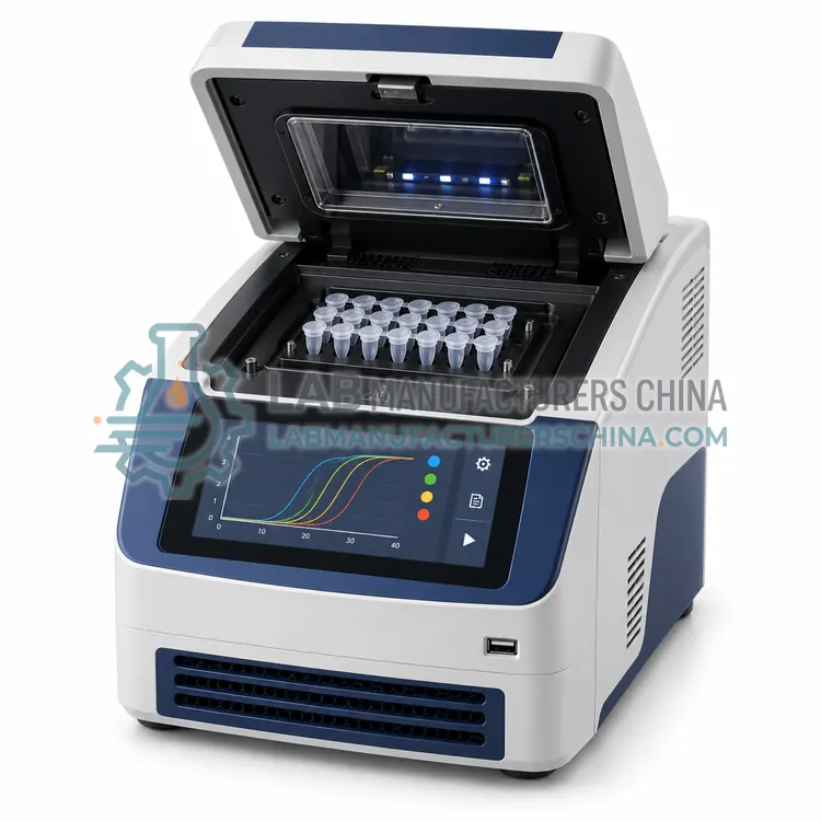
A benchtop flow cytometer includes a fluidics system with sheath fluid that hydrodynamically focuses a sample stream, one to three lasers such as 405, 488 and 640 nm, collection optics with dichroic and bandpass filters, and detectors that convert light into pulses. Forward scatter relates to cell size, side scatter to granularity, and fluorescence channels detect labelled antibodies or dyes. Data acquisition rates reach 10,000 events per second or more. Analysis software supports gating, compensation for spectral overlap, statistics and batch processing. Systems use either pressure-driven or syringe-driven fluidics, the latter allowing absolute counts without beads. Daily quality control with calibration beads, sheath fluid supply and waste decontamination are routine. Compact designs fit on a standard bench.
How does it work?
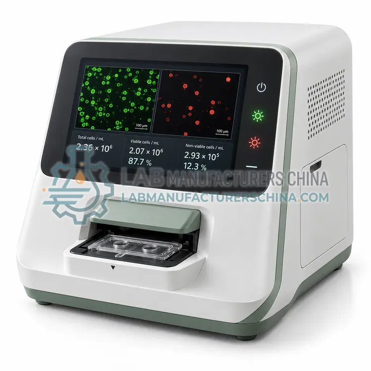
Cell analysis instruments use three main detection principles. Image-based counters photograph cells in a counting chamber, then software segments cells and classifies them as live or dead by dye exclusion, for example trypan blue, or by fluorescence with acridine orange and propidium iodide. Impedance counters, based on the Coulter principle, draw cells through a micro-aperture and record the electrical resistance pulse, whose amplitude is proportional to cell volume. Flow cytometers hydrodynamically focus cells into a single file past one or more lasers, detect forward and side scatter plus fluorescence through dichroic mirrors and bandpass filters, and convert photons to digital events with photomultiplier tubes or avalanche photodiodes. Software then gates populations and reports concentration, viability, size distribution and marker expression.
What are the key features?
- One to three lasers (405, 488, 640 nm) with 2 to 8 fluorescence channels.
- Hydrodynamic focusing with sheath fluid for stable event detection.
- Acquisition up to 10,000 events per second or more.
- Compensation and gating software with batch analysis.
- Syringe-driven option provides absolute counts without beads.
- Automated startup, shutdown and cleaning routines.
What are the technical specifications?
A typical benchtop flow cytometer configuration offers lasers of 488 nm, optional 405 and 640 nm, fluorescence channels of 2–8 colors, scatter detection of FSC, SSC, acquisition rate of up to 10,000–35,000 events/s, sample flow rate of 10–120 μL/min and fluorescence sensitivity of FITC below 500 MESF (typical). Values are indicative of the product class; the exact figures for each configuration are confirmed on the quotation and the calibration documents supplied with the instrument.
Which applications is it used for?
The Benchtop Flow Cytometer is used in immunophenotyping of lymphocyte subsets, apoptosis and cell cycle analysis, transfection efficiency with fluorescent proteins and microbial and bead-based assay analysis. Across the wider Cell Analysis class, laboratories also deploy it for cell culture seeding and passaging, cell therapy product characterization and bioprocess viability monitoring.
Which standards and certifications apply?
Relevant standards and methods include IEC 61010-1, IEC 61010-2-101, IEC 61326-2-6, IEC 60825-1, ISO 13322-1 and ISO 13319. Certification candidates for this model are CE, ISO 9001 manufacturing and IEC 60825-1 laser product classification; each is to be confirmed for the specific configuration before shipment, and certificates are supplied only where they have been issued.
Cell analyzers need regular optical and fluidic verification. Image counters should be checked with count-bead or latex standards, and impedance counters with calibration particles at the aperture size. Flow cytometers require daily quality control with fluorescent beads, fluidic cleaning after each session and periodic laser alignment checks. Install flow cytometers away from vibration and heat, with stable power and, if needed, a waste decontamination procedure for biohazardous samples. Use consumables from the validated lot, handle cell suspensions gently to avoid clumping, and standardize dye concentrations and incubation times. Regulated laboratories should verify software access control and audit trail.
How do I select the right Benchtop Flow Cytometer?
Select a benchtop cytometer when multi-parameter single-cell data are required but sorting is not. Specify the lasers and channels that match your fluorochromes, acquisition speed, plate loader, absolute-count capability and software access control. For sorting or more than eight colors, consider larger cell-sorting platforms. Request demonstration data on your own samples before purchase.
How does it compare with other Cell Analysis variants?
Start with the question you need answered. For concentration and viability only, an image-based counter or impedance counter is fast and inexpensive. For immunophenotyping, cell cycle or apoptosis, you need a flow cytometer with the right lasers and detector channels for your fluorochromes. Consider sample characteristics: clumping, debris, primary cells or platelets affect image segmentation, and small particles require an impedance aperture of suitable diameter. Check the counting range in cells per mL and the sample volume required, especially for scarce samples. Review consumable costs, since slides or cartridges often dominate running costs. Confirm software features for data export, user control and audit trail, and whether the instrument supports the sample types used in cell therapy or bioprocess workflows.
Full specifications
| Lasers | 488 nm, optional 405 and 640 nm |
|---|---|
| Fluorescence channels | 2–8 colors |
| Scatter detection | FSC, SSC |
| Acquisition rate | up to 10,000–35,000 events/s |
| Sample flow rate | 10–120 μL/min |
| Fluorescence sensitivity | FITC below 500 MESF (typical) |
| Particle size range | 0.5–40 μm |
| Sample tube format | 12 × 75 mm tubes, optional plate loader |
| Sheath fluid consumption | approx. 3–10 mL/h |
| Power supply | 100–240 V, 50/60 Hz |
Indicative values for this product class. Final specifications are confirmed on the quotation and in the calibration documents supplied with the instrument.
Applications
- Immunophenotyping of lymphocyte subsets
- Apoptosis and cell cycle analysis
- Transfection efficiency with fluorescent proteins
- Microbial and bead-based assay analysis
Key features
- One to three lasers (405, 488, 640 nm) with 2 to 8 fluorescence channels.
- Hydrodynamic focusing with sheath fluid for stable event detection.
- Acquisition up to 10,000 events per second or more.
- Compensation and gating software with batch analysis.
- Syringe-driven option provides absolute counts without beads.
- Automated startup, shutdown and cleaning routines.
Frequently asked questions about Benchtop Flow Cytometer
Match lasers to your fluorochromes. A 488 nm laser supports FITC, PE and PerCP dyes, a 640 nm laser adds APC and far-red dyes, and a 405 nm laser supports violet dyes and viability stains. Plan panels before selecting configuration.
Fluorochromes emit over broad wavelength ranges, so signal from one dye leaks into neighboring detectors. Compensation uses single-stained controls to subtract this spillover mathematically, which prevents false positive populations and ensures accurate quantification in multicolor panels.
Standard benchtop analyzers do not sort. Cell sorters use a charged droplet or microfluidic mechanism to physically separate populations and require additional biosafety and maintenance. If sorting is needed, specify a sorter or use a core facility.
A cell analyzer measures how many cells are in a sample, what fraction are alive, their size distribution and, with fluorescence, which markers they express. Laboratories use it to standardize seeding density, monitor culture health, check cell therapy products and characterize immune populations.
Automated counters typically deliver lower operator variability, with coefficients of variation of about 5 to 10 percent versus 15 to 25 percent for manual counting, but accuracy depends on focus, cell morphology and dye handling. Comparing results with a reference method during installation is good practice.
Trypan blue is excluded by live cells and stains dead cells blue, suitable for brightfield imaging. AO/PI uses fluorescence: acridine orange labels all nucleated cells and propidium iodide labels dead cells. AO/PI works better for samples with debris or red blood cells and in low-concentration samples.
Related models