Image-Based Automated Cell Counter
图像式自动细胞计数仪
- SKU CEA-0001
- Cell Analysis
- CE (to be confirmed)
- ISO 9001 manufacturing (to be confirmed)
Price on request — FOB / CIF quoted per order
Prices depend on configuration, quantity and destination port.
Key specifications
| Counting range | 1 × 10⁴–1 × 10⁷ cells/mL |
|---|---|
| Cell size range | 5–60 μm |
| Sample volume | 10 μL |
| Time per count | 10–30 s |
| Viability method | Trypan blue exclusion |
| Camera | 5 MP CMOS |
Request a quote Download brochure · to be provided
By Lab Manufacturers China Technical TeamLast reviewed Standards: IEC 61010-1, IEC 61010-2-101, IEC 61326-2-6
What is the Image-Based Automated Cell Counter?
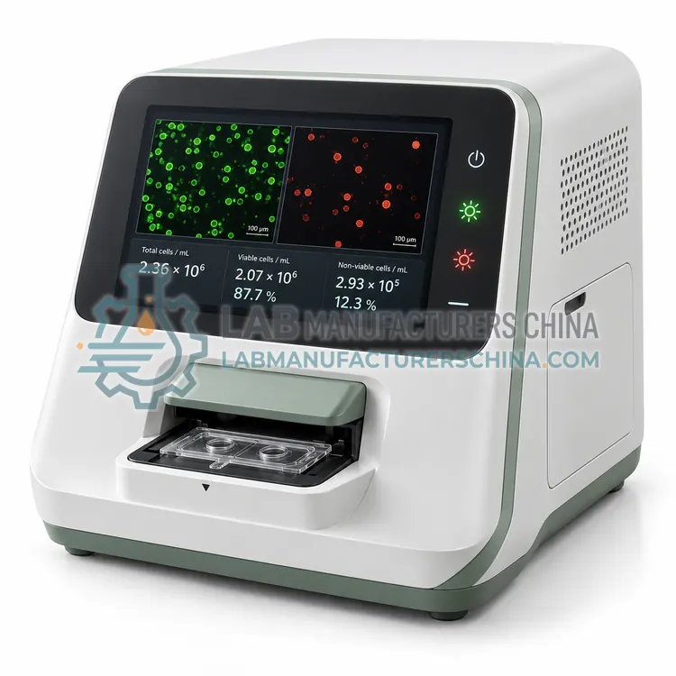
An image-based automated cell counter photographs a stained cell suspension in a disposable slide and uses software to count cells and classify viability by trypan blue exclusion. It suits routine culture laboratories needing results in under a minute from 10 μL of sample.
What is the Image-Based Automated Cell Counter?
An image-based automated cell counter photographs a stained cell suspension in a disposable slide and uses software to count cells and classify viability by trypan blue exclusion. It suits routine culture laboratories needing results in under a minute from 10 μL of sample.
Overview
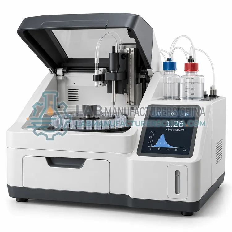
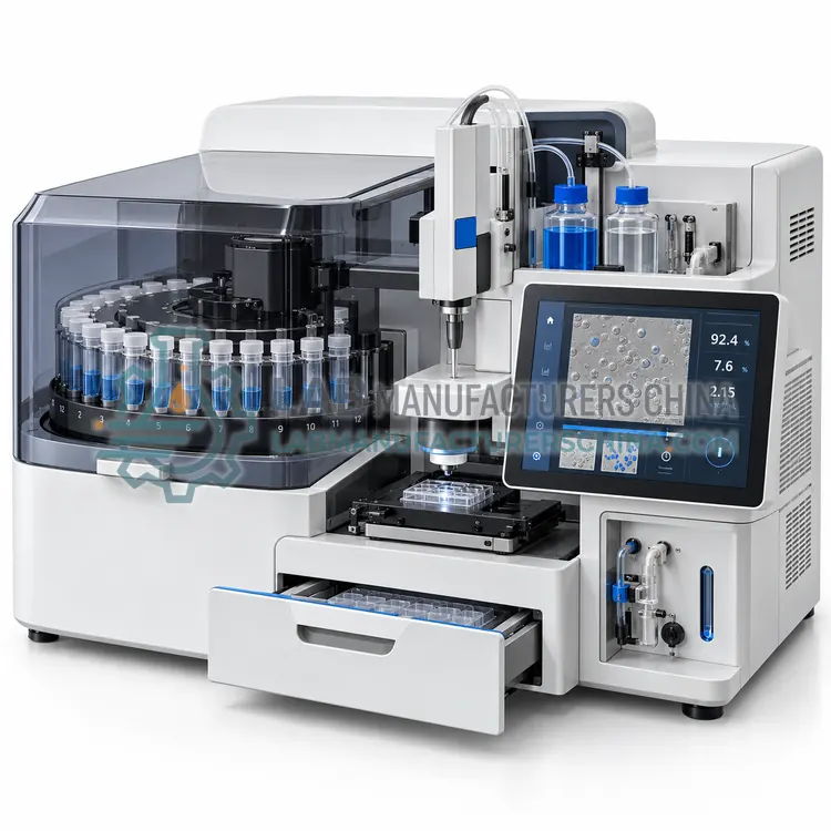
This counter combines a brightfield optical module, a CMOS camera and image-analysis algorithms. A 10 μL sample mixed 1:1 with trypan blue is loaded into a disposable counting slide with two chambers, and the instrument autofocuses, captures one or several fields, and identifies cells by size, circularity and intensity. Live cells appear bright and dead cells dark, allowing the software to report total and live concentration, viability percent, average diameter and aggregate fraction. Typical counting range is 1 × 10⁴ to 1 × 10⁷ cells/mL, extendable by dilution. Adjustable size gates and focus settings adapt the counter to different cell lines, while image storage allows visual review of counts. Results are available in about 10 to 30 seconds per sample.
How does it work?
Cell analysis instruments use three main detection principles. Image-based counters photograph cells in a counting chamber, then software segments cells and classifies them as live or dead by dye exclusion, for example trypan blue, or by fluorescence with acridine orange and propidium iodide. Impedance counters, based on the Coulter principle, draw cells through a micro-aperture and record the electrical resistance pulse, whose amplitude is proportional to cell volume. Flow cytometers hydrodynamically focus cells into a single file past one or more lasers, detect forward and side scatter plus fluorescence through dichroic mirrors and bandpass filters, and convert photons to digital events with photomultiplier tubes or avalanche photodiodes. Software then gates populations and reports concentration, viability, size distribution and marker expression.
What are the key features?
- Autofocus brightfield imaging with disposable dual-chamber counting slides.
- Reports concentration, viability, mean diameter and aggregate ratio.
- Uses only 10 μL of sample mixed with trypan blue.
- Adjustable cell-size gates for different cell lines.
- Stores images and results for review and export via USB or network.
- Compact footprint suits cell culture hood-side use.
What are the technical specifications?
A typical image-based automated cell counter configuration offers counting range of 1 × 10⁴–1 × 10⁷ cells/mL, cell size range of 5–60 μm, sample volume of 10 μL, time per count of 10–30 s, viability method of Trypan blue exclusion and camera of 5 MP CMOS. Values are indicative of the product class; the exact figures for each configuration are confirmed on the quotation and the calibration documents supplied with the instrument.
Which applications is it used for?
The Image-Based Automated Cell Counter is used in routine cell culture passaging, seeding density standardization, transfection and cell assay preparation and teaching laboratories. Across the wider Cell Analysis class, laboratories also deploy it for cell culture seeding and passaging, cell therapy product characterization and bioprocess viability monitoring.
Which standards and certifications apply?
Relevant standards and methods include IEC 61010-1, IEC 61010-2-101, IEC 61326-2-6, IEC 60825-1, ISO 13322-1 and ISO 13319. Certification candidates for this model are CE and ISO 9001 manufacturing; each is to be confirmed for the specific configuration before shipment, and certificates are supplied only where they have been issued.
Cell analyzers need regular optical and fluidic verification. Image counters should be checked with count-bead or latex standards, and impedance counters with calibration particles at the aperture size. Flow cytometers require daily quality control with fluorescent beads, fluidic cleaning after each session and periodic laser alignment checks. Install flow cytometers away from vibration and heat, with stable power and, if needed, a waste decontamination procedure for biohazardous samples. Use consumables from the validated lot, handle cell suspensions gently to avoid clumping, and standardize dye concentrations and incubation times. Regulated laboratories should verify software access control and audit trail.
How do I select the right Image-Based Automated Cell Counter?
Select this variant for routine counting of cultured cells where trypan blue viability is sufficient and operators change often. Specify counting range, cell size range, slide consumables, data export format and 21 CFR Part 11 needs. Choose a fluorescence counter for debris-rich or primary samples.
How does it compare with other Cell Analysis variants?
Start with the question you need answered. For concentration and viability only, an image-based counter or impedance counter is fast and inexpensive. For immunophenotyping, cell cycle or apoptosis, you need a flow cytometer with the right lasers and detector channels for your fluorochromes. Consider sample characteristics: clumping, debris, primary cells or platelets affect image segmentation, and small particles require an impedance aperture of suitable diameter. Check the counting range in cells per mL and the sample volume required, especially for scarce samples. Review consumable costs, since slides or cartridges often dominate running costs. Confirm software features for data export, user control and audit trail, and whether the instrument supports the sample types used in cell therapy or bioprocess workflows.
Full specifications
| Counting range | 1 × 10⁴–1 × 10⁷ cells/mL |
|---|---|
| Cell size range | 5–60 μm |
| Sample volume | 10 μL |
| Time per count | 10–30 s |
| Viability method | Trypan blue exclusion |
| Camera | 5 MP CMOS |
| Counting precision (CV) | below 8 % (typical) |
| Display | 7-inch color touchscreen |
| Data export | USB, Ethernet, CSV/PDF |
| Power supply | 100–240 V, 50/60 Hz |
Indicative values for this product class. Final specifications are confirmed on the quotation and in the calibration documents supplied with the instrument.
Applications
- Routine cell culture passaging
- Seeding density standardization
- Transfection and cell assay preparation
- Teaching laboratories
Key features
- Autofocus brightfield imaging with disposable dual-chamber counting slides.
- Reports concentration, viability, mean diameter and aggregate ratio.
- Uses only 10 μL of sample mixed with trypan blue.
- Adjustable cell-size gates for different cell lines.
- Stores images and results for review and export via USB or network.
- Compact footprint suits cell culture hood-side use.
Frequently asked questions about Image-Based Automated Cell Counter
The best precision is achieved between about 2 × 10⁵ and 5 × 10⁶ cells/mL. Below this range, too few cells are captured per field, and above it, cell overlap increases error. Dilute or concentrate the sample to fall within the stated range.
It reports an aggregate ratio and can split small clusters using algorithms, but heavy clumping still underestimates counts. Dissociate cells thoroughly by pipetting or enzyme treatment, filter if necessary, and mix carefully before loading the slide.
Most image-based counters are designed around disposable dual-chamber slides that set a fixed depth, which is critical for accurate concentration. Reusable glass chambers are available on some models but require careful cleaning and depth verification.
A cell analyzer measures how many cells are in a sample, what fraction are alive, their size distribution and, with fluorescence, which markers they express. Laboratories use it to standardize seeding density, monitor culture health, check cell therapy products and characterize immune populations.
Automated counters typically deliver lower operator variability, with coefficients of variation of about 5 to 10 percent versus 15 to 25 percent for manual counting, but accuracy depends on focus, cell morphology and dye handling. Comparing results with a reference method during installation is good practice.
Trypan blue is excluded by live cells and stains dead cells blue, suitable for brightfield imaging. AO/PI uses fluorescence: acridine orange labels all nucleated cells and propidium iodide labels dead cells. AO/PI works better for samples with debris or red blood cells and in low-concentration samples.