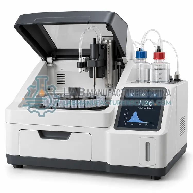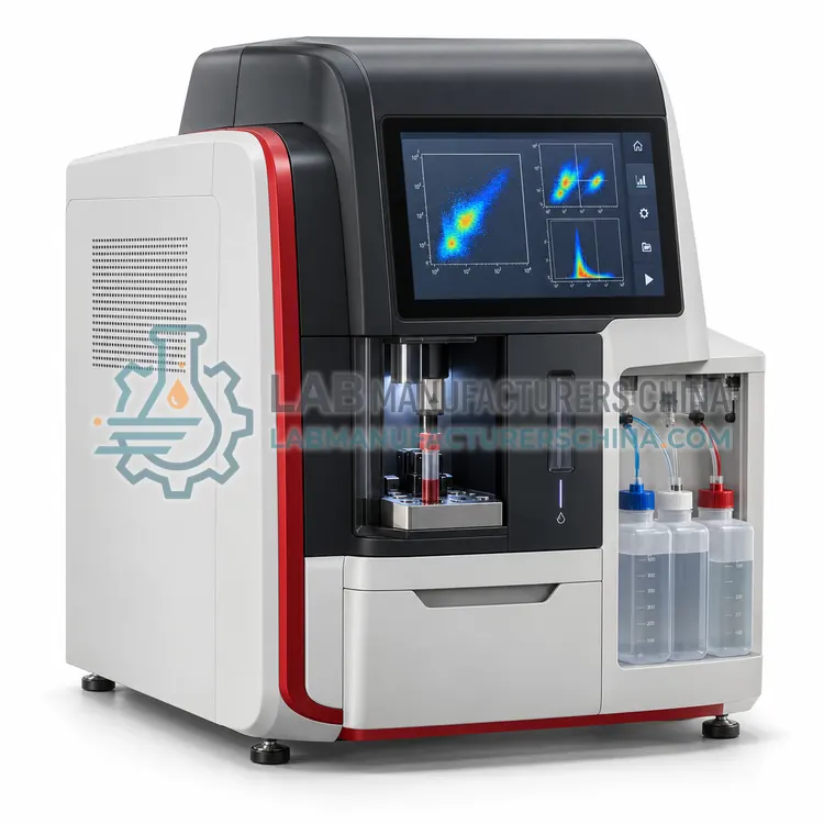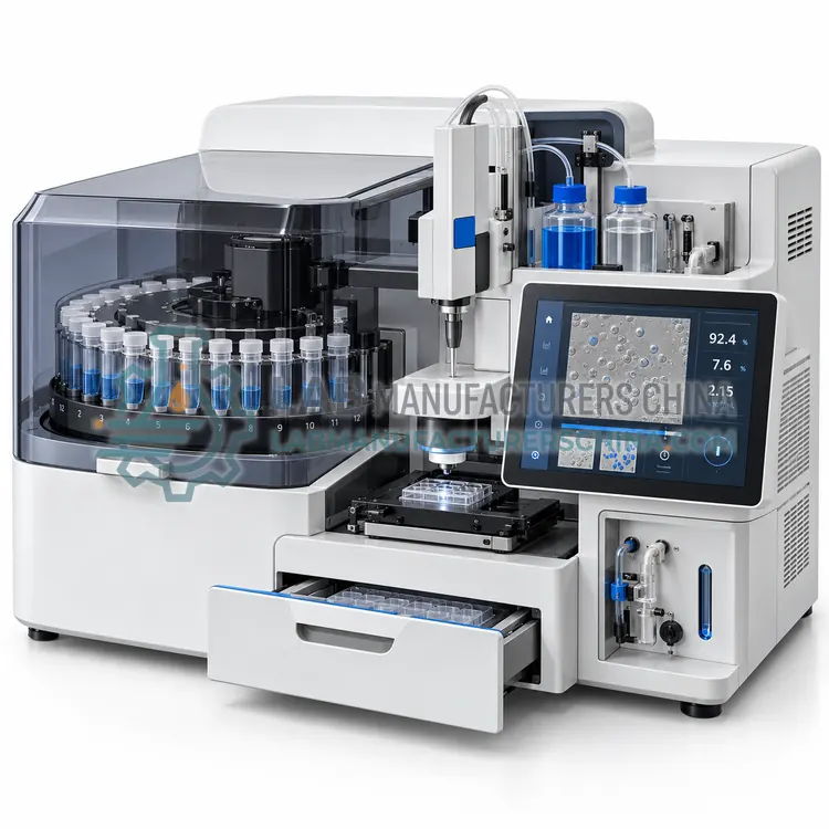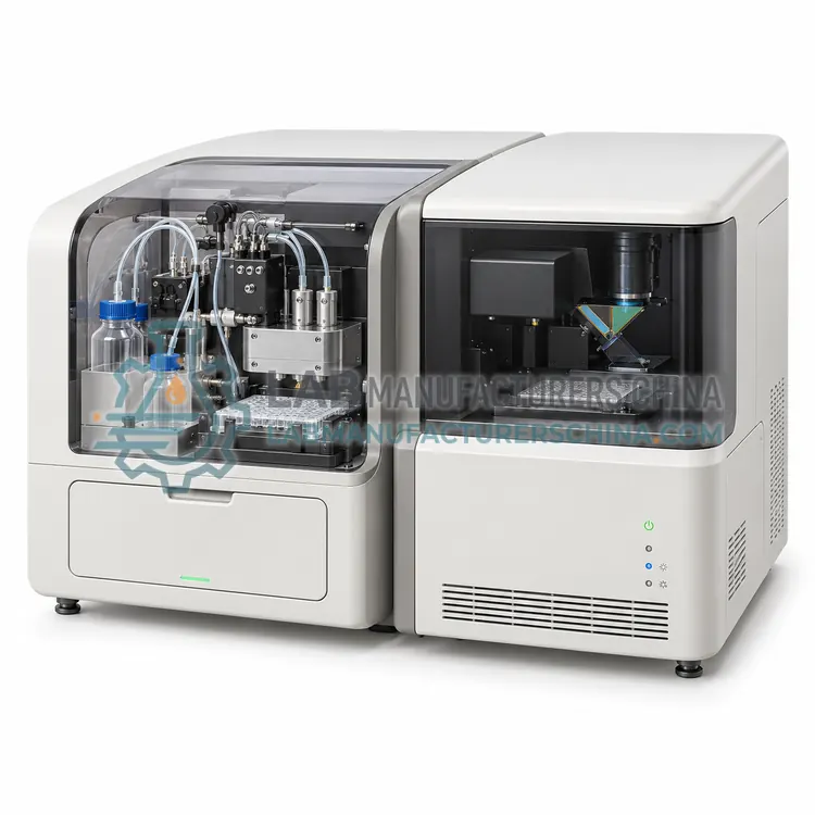
Fluorescence Cell Counter and Viability Analyzer
荧光细胞计数与活率分析仪
- SKU CEA-0002
- Cell Analysis
- CE (to be confirmed)
- ISO 9001 manufacturing (to be confirmed)
Price on request — FOB / CIF quoted per order
Prices depend on configuration, quantity and destination port.
Key specifications
| Counting range | 5 × 10⁴–3 × 10⁷ cells/mL |
|---|---|
| Fluorescence channels | 2 (green, red) |
| Excitation sources | Blue and green LEDs |
| Sample volume | 20 μL |
| Analysis time | approx. 30 s |
| Viability stains | AO/PI; trypan blue in brightfield |
Request a quote Download brochure · to be provided
By Lab Manufacturers China Technical TeamLast reviewed Standards: IEC 61010-1, IEC 61010-2-101, IEC 61326-2-6
What is the Fluorescence Cell Counter and Viability Analyzer?
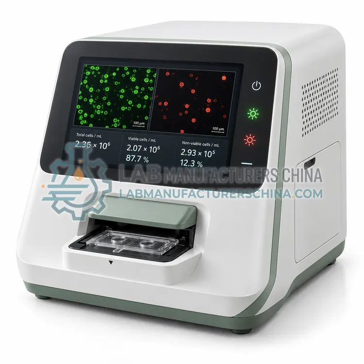
A fluorescence cell counter and viability analyzer uses acridine orange and propidium iodide staining with fluorescence imaging to count nucleated cells and determine viability. It is the better choice for primary cells, PBMCs and debris-rich samples where brightfield trypan blue methods are unreliable.
What is the Fluorescence Cell Counter and Viability Analyzer?
A fluorescence cell counter and viability analyzer uses acridine orange and propidium iodide staining with fluorescence imaging to count nucleated cells and determine viability. It is the better choice for primary cells, PBMCs and debris-rich samples where brightfield trypan blue methods are unreliable.
Overview
Fluorescence counters add excitation LEDs and emission filters to the image-based platform, typically a blue channel for acridine orange (green emission) and a green channel for propidium iodide (red emission). Acridine orange permeates all nucleated cells, whereas propidium iodide enters only cells with compromised membranes, so live and dead cells are identified by color. Because non-nucleated particles such as red blood cells, platelets and debris do not fluoresce, the method can count nucleated cells in whole blood or bone marrow without lysis. Counting range is typically 5 × 10⁴ to 3 × 10⁷ cells/mL, and analysis takes about 30 seconds. Some models also provide brightfield mode for trypan blue. Fluorescence requires consumables and light-shielded optics, and LED lifetime should be specified.
How does it work?
Cell analysis instruments use three main detection principles. Image-based counters photograph cells in a counting chamber, then software segments cells and classifies them as live or dead by dye exclusion, for example trypan blue, or by fluorescence with acridine orange and propidium iodide. Impedance counters, based on the Coulter principle, draw cells through a micro-aperture and record the electrical resistance pulse, whose amplitude is proportional to cell volume. Flow cytometers hydrodynamically focus cells into a single file past one or more lasers, detect forward and side scatter plus fluorescence through dichroic mirrors and bandpass filters, and convert photons to digital events with photomultiplier tubes or avalanche photodiodes. Software then gates populations and reports concentration, viability, size distribution and marker expression.
What are the key features?
- Dual fluorescence channels for acridine orange and propidium iodide.
- Counts nucleated cells without interference from red cells and debris.
- Optional brightfield mode for trypan blue assays.
- Autofocus and multi-field imaging improve counting statistics.
- Validated assay protocols for PBMC, bone marrow and cell lines.
- Image archive and audit-trail-capable software options.
What are the technical specifications?
A typical fluorescence cell counter and viability analyzer configuration offers counting range of 5 × 10⁴–3 × 10⁷ cells/mL, fluorescence channels of 2 (green, red), excitation sources of Blue and green LEDs, sample volume of 20 μL, analysis time of approx. 30 s and viability stains of AO/PI; trypan blue in brightfield. Values are indicative of the product class; the exact figures for each configuration are confirmed on the quotation and the calibration documents supplied with the instrument.
Which applications is it used for?
The Fluorescence Cell Counter and Viability Analyzer is used in PBMC and lymphocyte isolation QC, cell therapy product release testing, bone marrow and whole blood nucleated cell counts and primary cell culture. Across the wider Cell Analysis class, laboratories also deploy it for cell culture seeding and passaging, cell therapy product characterization and bioprocess viability monitoring.
Which standards and certifications apply?
Relevant standards and methods include IEC 61010-1, IEC 61010-2-101, IEC 61326-2-6, IEC 60825-1, ISO 13322-1 and ISO 13319. Certification candidates for this model are CE and ISO 9001 manufacturing; each is to be confirmed for the specific configuration before shipment, and certificates are supplied only where they have been issued.
Cell analyzers need regular optical and fluidic verification. Image counters should be checked with count-bead or latex standards, and impedance counters with calibration particles at the aperture size. Flow cytometers require daily quality control with fluorescent beads, fluidic cleaning after each session and periodic laser alignment checks. Install flow cytometers away from vibration and heat, with stable power and, if needed, a waste decontamination procedure for biohazardous samples. Use consumables from the validated lot, handle cell suspensions gently to avoid clumping, and standardize dye concentrations and incubation times. Regulated laboratories should verify software access control and audit trail.
How do I select the right Fluorescence Cell Counter and Viability Analyzer?
Choose this variant when samples contain red cells, platelets or debris, or when low cell concentrations require fluorescence sensitivity. Specify channels, stain chemistry, consumables, validation documents and software control features. For clean cell line cultures, a brightfield counter is often sufficient and cheaper. Request demonstration data on your own samples before purchase.
How does it compare with other Cell Analysis variants?
Start with the question you need answered. For concentration and viability only, an image-based counter or impedance counter is fast and inexpensive. For immunophenotyping, cell cycle or apoptosis, you need a flow cytometer with the right lasers and detector channels for your fluorochromes. Consider sample characteristics: clumping, debris, primary cells or platelets affect image segmentation, and small particles require an impedance aperture of suitable diameter. Check the counting range in cells per mL and the sample volume required, especially for scarce samples. Review consumable costs, since slides or cartridges often dominate running costs. Confirm software features for data export, user control and audit trail, and whether the instrument supports the sample types used in cell therapy or bioprocess workflows.
Full specifications
| Counting range | 5 × 10⁴–3 × 10⁷ cells/mL |
|---|---|
| Fluorescence channels | 2 (green, red) |
| Excitation sources | Blue and green LEDs |
| Sample volume | 20 μL |
| Analysis time | approx. 30 s |
| Viability stains | AO/PI; trypan blue in brightfield |
| Cell size range | 4–60 μm |
| Precision (CV) | below 6 % (typical) |
| Data export | USB, LIMS-ready CSV |
| Power supply | 100–240 V, 50/60 Hz |
Indicative values for this product class. Final specifications are confirmed on the quotation and in the calibration documents supplied with the instrument.
Applications
- PBMC and lymphocyte isolation QC
- Cell therapy product release testing
- Bone marrow and whole blood nucleated cell counts
- Primary cell culture
Key features
- Dual fluorescence channels for acridine orange and propidium iodide.
- Counts nucleated cells without interference from red cells and debris.
- Optional brightfield mode for trypan blue assays.
- Autofocus and multi-field imaging improve counting statistics.
- Validated assay protocols for PBMC, bone marrow and cell lines.
- Image archive and audit-trail-capable software options.
Frequently asked questions about Fluorescence Cell Counter and Viability Analyzer
Acridine orange and propidium iodide stain only nucleated cells, so platelets, debris and residual red cells do not distort counts. Trypan blue stains any dead particle and requires subjective judgment in debris-rich samples, leading to viability overestimation or underestimation.
Propidium iodide and acridine orange are potential mutagens. Use gloves and eye protection, collect stained waste separately according to local chemical waste rules, and follow supplier safety data sheets. Pre-mixed dye solutions reduce handling exposure.
Calibration uses fluorescent count beads or reference cell standards at known concentrations to verify intensity, focus and counting accuracy. Laboratories typically check monthly or after service, and the manufacturer may supply a calibration or verification kit.
A cell analyzer measures how many cells are in a sample, what fraction are alive, their size distribution and, with fluorescence, which markers they express. Laboratories use it to standardize seeding density, monitor culture health, check cell therapy products and characterize immune populations.
Automated counters typically deliver lower operator variability, with coefficients of variation of about 5 to 10 percent versus 15 to 25 percent for manual counting, but accuracy depends on focus, cell morphology and dye handling. Comparing results with a reference method during installation is good practice.
Trypan blue is excluded by live cells and stains dead cells blue, suitable for brightfield imaging. AO/PI uses fluorescence: acridine orange labels all nucleated cells and propidium iodide labels dead cells. AO/PI works better for samples with debris or red blood cells and in low-concentration samples.
Related models