How do Immunoblot Systems work?
Immunoblotting starts with proteins that are either separated by polyacrylamide gel electrophoresis and transferred to a membrane, or applied directly as discrete lines on a strip. The membrane is blocked to prevent non-specific binding, then incubated with diluted patient sample so that antibodies in the sample bind to their target antigens. After washing, an enzyme- or fluorophore-labelled anti-human antibody is added, and a substrate produces a visible, luminescent or fluorescent signal proportional to the bound antibody. Strips are read by eye, by a scanning densitometer or by a charge-coupled device camera, and each band is scored as absent, weak, moderate or strong, or quantified as an intensity ratio against a control band. Interpretation criteria for confirmatory assays are set by the manufacturer and by national guidance, and the pattern of reactive bands, not a single band, determines the reported result.
Which Immunoblot Systems variants are available?
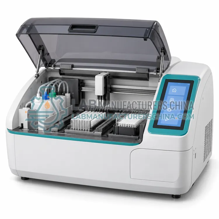
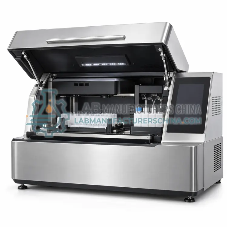
- Automated Immunoblot Processor — An automated immunoblot processor performs the incubation, washing and conjugate steps for membrane strips or blots under program control, then presents them for reading.
- Semi-Automated Line Immunoassay Strip Processor — This instrument automates reagent dispensing, rocking and washing for line immunoassay strips while the operator handles sample addition and transfer between trays.
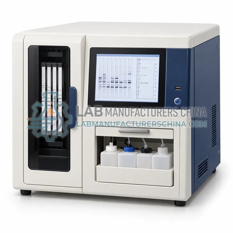
- Immunoblot Strip Reader and Densitometer — This instrument scans stained or luminescent immunoblot strips, measures band intensity by densitometry and applies the manufacturer's interpretation criteria automatically.
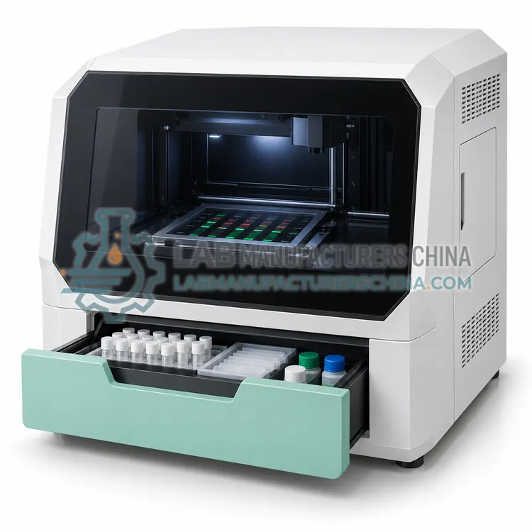
- Chemiluminescence Western Blot Imaging System — This imaging system captures the light emitted by chemiluminescent Western blot membranes using a cooled charge-coupled device camera and quantifies band intensity without film.
- Fluorescence Western Blot Imaging System — This system excites fluorescently labelled antibodies on a blot and captures two or more channels in one image, allowing multiplex detection and total protein normalization.
How do I choose the right variant?
Decide first whether the laboratory processes strips manually or needs automated incubation and washing, because the processing step dominates hands-on time and reproducibility. Then choose the reading method: visual reading suits low volumes and is accepted for many line immunoassays, densitometry provides objective band intensity, and camera-based imaging supports chemiluminescence and multiplex fluorescence. Check that the instrument accepts the strip and membrane formats of your kits, that the software applies the manufacturer interpretation criteria, and that images and results are archived in a retrievable format. Confirm assay registration in your market, and the availability of controls, conjugates and substrates, since these consumables are kit specific and may have short shelf lives.
Where are Immunoblot Systems used?
Immunoblot Systems are specified in HIV and hepatitis confirmatory testing, autoimmune antibody profile testing, lyme disease and syphilis confirmation and allergen component and research blotting.
Which standards and certifications apply?
Work with Immunoblot Systems is commonly governed by ISO 15189, CLSI M53, CLSI EP05, CLSI EP17, WHO HIV testing strategy, IEC 61010-1 and IEC 61010-2-101. Instrument certifications such as CE marking are confirmed per configuration and never assumed.
What are the key considerations before purchase?
Work on a clean bench with gloves, since membranes are easily contaminated, and use only the buffers and dilutions specified in the kit, because salt concentration and pH change binding and washing efficiency. Keep incubation temperatures and times consistent, as band intensity varies with both, and avoid letting strips dry out between steps. Verify performance at installation with the kit's positive and negative controls and with a proficiency panel if available, and repeat the check with each new reagent lot. Clean the scanner or imaging optics regularly, keep the light source stable for fluorescence and luminescence work, and store images together with the patient identifier for the retention period required by local rules.