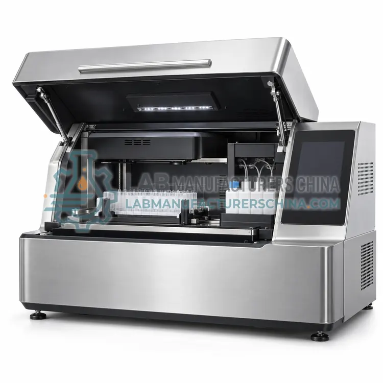
Chemiluminescence Western Blot Imaging System
化学发光 Western Blot 成像系统
- SKU IMU-0004
- Immunoblot Systems
- CE (to be confirmed)
- ISO 13485 manufacturing (to be confirmed)
- NMPA registration for research use (to be confirmed)
Price on request — FOB / CIF quoted per order
Prices depend on configuration, quantity and destination port.
Key specifications
| Detector | Cooled CCD, −25 to −40 °C |
|---|---|
| Dynamic range | Typically 4 orders of magnitude |
| Exposure range | 0.1 s to 30 min programmable |
| Membrane sizes | Up to 20 × 20 cm |
| Light channels | Chemiluminescence, white light, optional fluorescence |
| Resolution | 1–5 megapixel images per exposure |
Request a quote Download brochure · to be provided
By Lab Manufacturers China Technical TeamLast reviewed Standards: ISO 15189, CLSI M53, CLSI EP05
What is the Chemiluminescence Western Blot Imaging System?
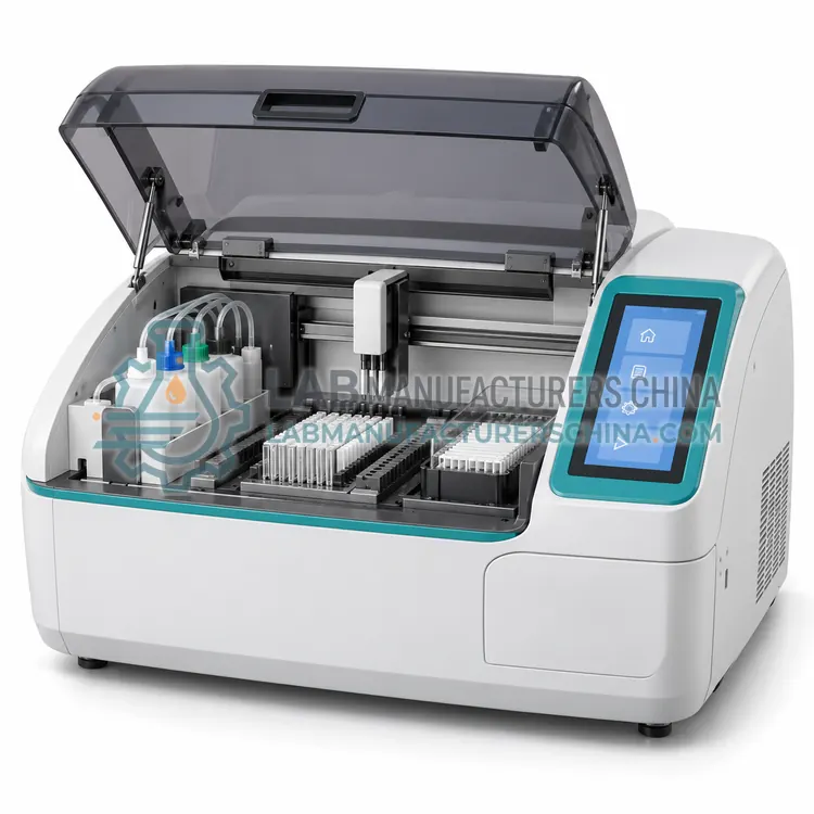
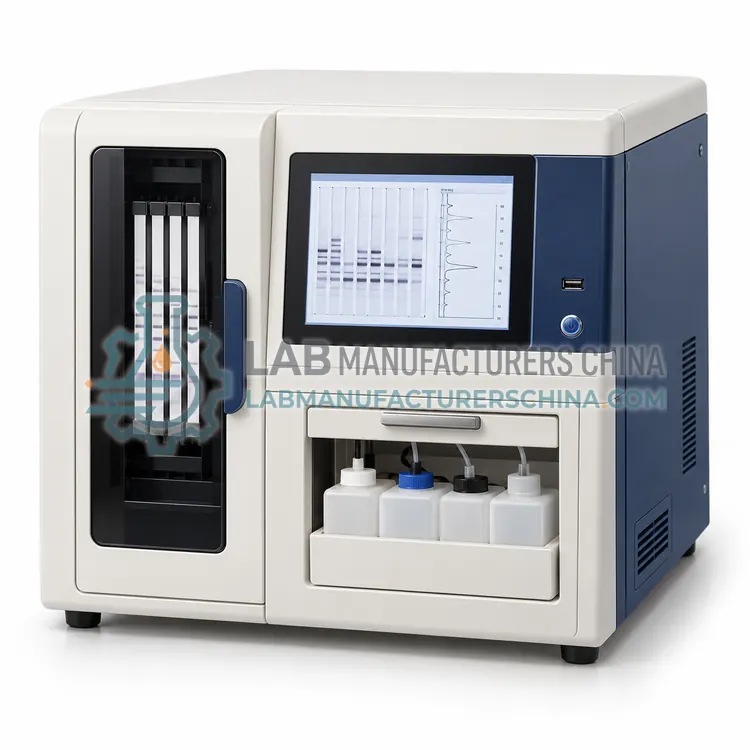
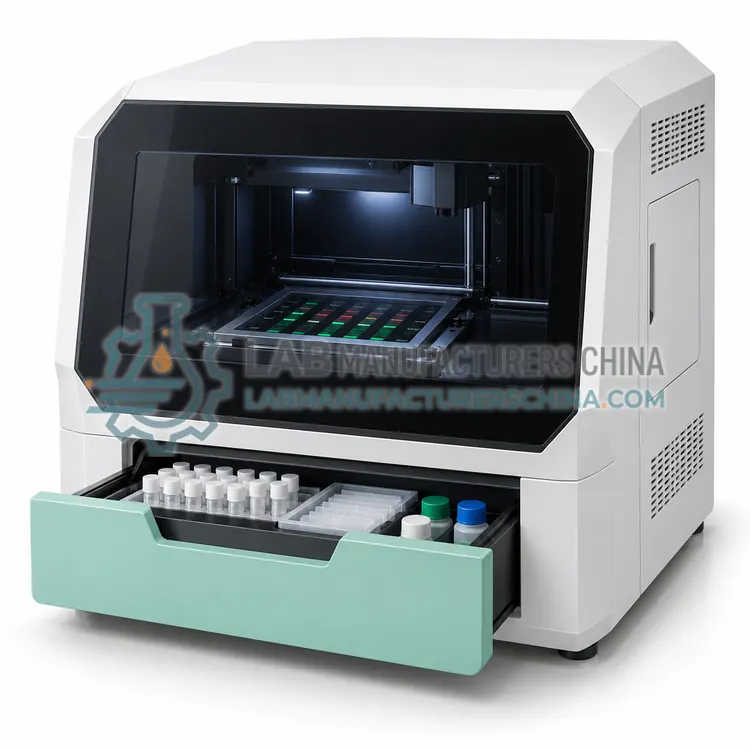
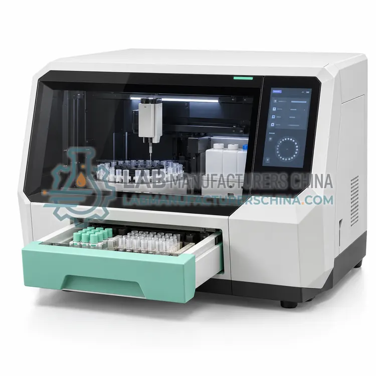
This imaging system captures the light emitted by chemiluminescent Western blot membranes using a cooled charge-coupled device camera and quantifies band intensity without film. It suits laboratories that need sensitive detection with a wide linear range and digital records.
What is the Chemiluminescence Western Blot Imaging System?
This imaging system captures the light emitted by chemiluminescent Western blot membranes using a cooled charge-coupled device camera and quantifies band intensity without film. It suits laboratories that need sensitive detection with a wide linear range and digital records.
Overview
The membrane is incubated with a horseradish peroxidase or alkaline phosphatase conjugate and a chemiluminescent substrate, and the light emitted where the conjugate has bound is captured by a cooled camera. Cooling reduces dark current so that long exposures produce low-noise images, and the software subtracts background, locates lanes and bands, and calculates normalized intensity against a loading control. Unlike film, the camera has a wider linear response, so saturated bands are less likely, and quantitative comparison across a membrane is more reliable. Multiple exposures can be taken from the same membrane to cover weak and strong bands, and the images can be stored with the associated analysis. Typical instruments support various membrane sizes and include white light or fluorescence channels for total protein normalization.
How does it work?
Immunoblotting starts with proteins that are either separated by polyacrylamide gel electrophoresis and transferred to a membrane, or applied directly as discrete lines on a strip. The membrane is blocked to prevent non-specific binding, then incubated with diluted patient sample so that antibodies in the sample bind to their target antigens. After washing, an enzyme- or fluorophore-labelled anti-human antibody is added, and a substrate produces a visible, luminescent or fluorescent signal proportional to the bound antibody. Strips are read by eye, by a scanning densitometer or by a charge-coupled device camera, and each band is scored as absent, weak, moderate or strong, or quantified as an intensity ratio against a control band. Interpretation criteria for confirmatory assays are set by the manufacturer and by national guidance, and the pattern of reactive bands, not a single band, determines the reported result.
What are the key features?
- Cooled charge-coupled device camera with low dark current
- Wide linear dynamic range compared with film detection
- Multiple exposure capture from one membrane
- Automated lane and band detection with background subtraction
- White light or fluorescence channels for total protein normalization
- Digital image archive with analysis parameters
What are the technical specifications?
A typical chemiluminescence western blot imaging system configuration offers detector of Cooled CCD, −25 to −40 °C, dynamic range of Typically 4 orders of magnitude, exposure range of 0.1 s to 30 min programmable, membrane sizes of Up to 20 × 20 cm, light channels of Chemiluminescence, white light, optional fluorescence and resolution of 1–5 megapixel images per exposure. Values are indicative of the product class; the exact figures for each configuration are confirmed on the quotation and the calibration documents supplied with the instrument.
Which applications is it used for?
The Chemiluminescence Western Blot Imaging System is used in western blot confirmatory testing, research protein expression studies, autoimmune blot confirmations and quality control of antigen preparations. Across the wider Immunoblot Systems class, laboratories also deploy it for HIV and hepatitis confirmatory testing, autoimmune antibody profile testing and lyme disease and syphilis confirmation.
Which standards and certifications apply?
Relevant standards and methods include ISO 15189, CLSI M53, CLSI EP05, CLSI EP17, WHO HIV testing strategy, IEC 61010-1 and IEC 61010-2-101. Certification candidates for this model are CE, ISO 13485 manufacturing and NMPA registration for research use; each is to be confirmed for the specific configuration before shipment, and certificates are supplied only where they have been issued.
Work on a clean bench with gloves, since membranes are easily contaminated, and use only the buffers and dilutions specified in the kit, because salt concentration and pH change binding and washing efficiency. Keep incubation temperatures and times consistent, as band intensity varies with both, and avoid letting strips dry out between steps. Verify performance at installation with the kit's positive and negative controls and with a proficiency panel if available, and repeat the check with each new reagent lot. Clean the scanner or imaging optics regularly, keep the light source stable for fluorescence and luminescence work, and store images together with the patient identifier for the retention period required by local rules.
How do I select the right Chemiluminescence Western Blot Imaging System?
Specify this imaging system when chemiluminescent detection is used, when film-based reading is being retired, or when quantitative band comparison across lanes is needed. In the RFQ confirm the detector cooling specification, achievable dynamic range, exposure control, and whether the software supports the normalization strategy your laboratory uses.
How does it compare with other Immunoblot Systems variants?
Decide first whether the laboratory processes strips manually or needs automated incubation and washing, because the processing step dominates hands-on time and reproducibility. Then choose the reading method: visual reading suits low volumes and is accepted for many line immunoassays, densitometry provides objective band intensity, and camera-based imaging supports chemiluminescence and multiplex fluorescence. Check that the instrument accepts the strip and membrane formats of your kits, that the software applies the manufacturer interpretation criteria, and that images and results are archived in a retrievable format. Confirm assay registration in your market, and the availability of controls, conjugates and substrates, since these consumables are kit specific and may have short shelf lives.
Full specifications
| Detector | Cooled CCD, −25 to −40 °C |
|---|---|
| Dynamic range | Typically 4 orders of magnitude |
| Exposure range | 0.1 s to 30 min programmable |
| Membrane sizes | Up to 20 × 20 cm |
| Light channels | Chemiluminescence, white light, optional fluorescence |
| Resolution | 1–5 megapixel images per exposure |
| Software functions | Background subtraction, lane and band quantitation |
| Interfaces | USB, LAN, network storage export |
Indicative values for this product class. Final specifications are confirmed on the quotation and in the calibration documents supplied with the instrument.
Applications
- Western blot confirmatory testing
- Research protein expression studies
- Autoimmune blot confirmations
- Quality control of antigen preparations
Key features
- Cooled charge-coupled device camera with low dark current
- Wide linear dynamic range compared with film detection
- Multiple exposure capture from one membrane
- Automated lane and band detection with background subtraction
- White light or fluorescence channels for total protein normalization
- Digital image archive with analysis parameters
Frequently asked questions about Chemiluminescence Western Blot Imaging System
Cooling reduces dark current, the background signal the detector produces without light, so long exposures remain low in noise. Without cooling, weak chemiluminescent bands would be lost in noise or would require signal averaging that lengthens the workflow.
Capture a series of exposures, for example ten seconds, one minute and five minutes, so that both weak and strong bands fall within the linear range. The software should record which exposure was used for each quantitation, since using a saturated exposure underestimates strong bands.
Yes, if it includes the appropriate excitation light sources and emission filters, which many systems offer as an option. Fluorescence enables multiplex detection and total protein normalization, and it requires attention to photobleaching and to the linearity of the fluorescence channel.
Yes, images should be stored with the analysis parameters, exposure times and reuse evidence for the retention period required locally. Retaining the raw image lets a reviewer re-analyse the data if a quantitative result is questioned after reporting, which is valuable for accreditation.
Western blot separates proteins by electrophoresis and transfers them to a membrane, so bands are positioned by molecular weight. Line immunoassay applies purified or recombinant antigens as discrete lines on a strip. Both are read by band pattern, and kits indicate which interpretation criteria apply.
It is accepted for many confirmatory assays when the kit instructions define scoring categories and the laboratory documents reader training and periodic competency assessment. Densitometry or imaging adds objectivity and an archived record, which many laboratories prefer for traceability and for resolving discrepant readings.
ISO 15189 covers laboratory quality, CLSI M53 addresses criteria for laboratory testing and diagnosis of HIV, and national programmes publish their own testing strategies. Precision and detection limit studies follow CLSI EP05 and EP17, and instrument electrical safety follows IEC 61010-1 and IEC 61010-2-101.
Related models