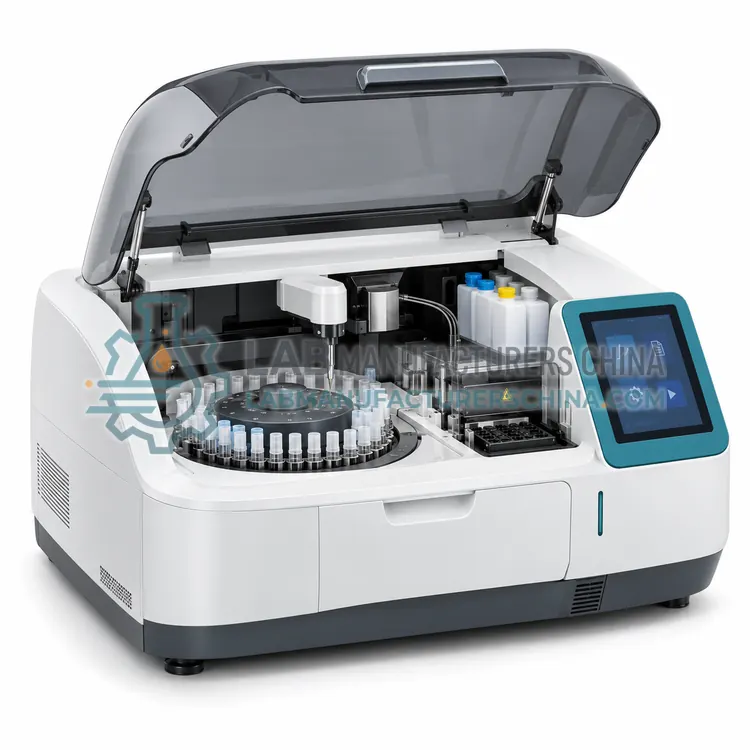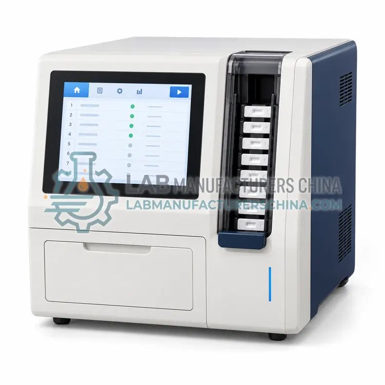
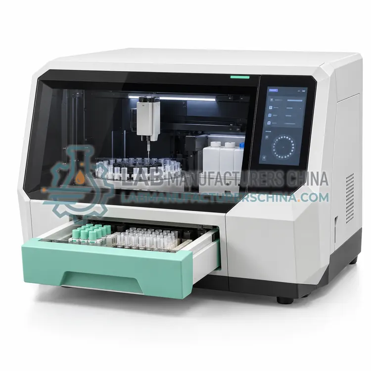
Fluorescence Polarization Immunoassay Analyzer
荧光偏振免疫分析仪
- SKU FLI-0005
- Fluorescence Immunoassay
- CE (to be confirmed)
- ISO 13485 manufacturing (to be confirmed)
- NMPA class II registration (to be confirmed)
Price on request — FOB / CIF quoted per order
Prices depend on configuration, quantity and destination port.
Key specifications
| Measurement principle | Fluorescence polarization of a labelled tracer |
|---|---|
| Excitation wavelength | Typically 480–500 nm |
| Emission wavelength | Typically 520–540 nm |
| Sample volume | 20–100 µL serum or plasma |
| Time to result | 2–6 min per analyte |
| Incubation temperature | 30 °C or 37 °C ±0.5 °C |
Request a quote Download brochure · to be provided
By Lab Manufacturers China Technical TeamLast reviewed Standards: ISO 15189, CLSI EP05, CLSI EP15
What is the Fluorescence Polarization Immunoassay Analyzer?
A fluorescence polarization immunoassay analyzer measures the change in polarization of fluorescently labelled small molecules as they bind to antibodies, giving a homogeneous assay with no separation step. It suits therapeutic drug monitoring and measurement of small analytes in serum.
What is the Fluorescence Polarization Immunoassay Analyzer?
A fluorescence polarization immunoassay analyzer measures the change in polarization of fluorescently labelled small molecules as they bind to antibodies, giving a homogeneous assay with no separation step. It suits therapeutic drug monitoring and measurement of small analytes in serum.
Overview
In fluorescence polarization the tracer is a small fluorescently labelled analogue of the analyte. Free tracer rotates rapidly in solution and emits light that is largely depolarized, while tracer bound to a much larger antibody rotates slowly and its emission retains the polarization of the excitation. Adding patient sample makes unlabelled analyte compete for antibody sites, so the free tracer fraction rises and polarization falls. The analyzer measures polarization directly, without washing or separation, which makes the assay fast and easy to automate and avoids errors from incomplete separation. The method suits small molecules such as therapeutic drugs, hormones and drugs of abuse, and results are available in a few minutes. Because the signal depends on binding equilibrium, temperature control and incubation timing are important, and each analyte needs a specific antibody and tracer.
How does it work?
Fluorescence immunoassay uses an antibody labelled with a fluorophore such as fluorescein, a europium chelate or a quantum dot. After the immune complex forms, the label is excited by light of one wavelength and emits light at a longer wavelength, and a photodetector measures the emission after optical filtering. The difference between excitation and emission wavelengths allows the instrument to separate the signal from the excitation light, and background fluorescence from sample components is reduced by careful filter selection, by washing away unbound label and, in time-resolved systems, by delaying the measurement until short-lived background fluorescence has decayed. Since the fluorescent signal is measured against a low background, sensitivity is generally better than absorbance-based enzyme immunoassay. Quantification relies on a calibration curve stored in the analyzer, and strip-based tests use a reader that scans the test and control lines.
What are the key features?
- Homogeneous assay with no washing or separation step
- Polarization measurement of tracer bound to antibody
- Results available within a few minutes
- Suitable for small molecules such as therapeutic drugs
- Temperature-controlled cuvette compartment for binding equilibrium
- Direct measurement reduces separation-related errors
What are the technical specifications?
A typical fluorescence polarization immunoassay analyzer configuration offers measurement principle of Fluorescence polarization of a labelled tracer, excitation wavelength of Typically 480–500 nm, emission wavelength of Typically 520–540 nm, sample volume of 20–100 µL serum or plasma, time to result of 2–6 min per analyte and incubation temperature of 30 °C or 37 °C ±0.5 °C. Values are indicative of the product class; the exact figures for each configuration are confirmed on the quotation and the calibration documents supplied with the instrument.
Which applications is it used for?
The Fluorescence Polarization Immunoassay Analyzer is used in therapeutic drug monitoring, drug of abuse screening in serum, small hormone analyte measurement and veterinary therapeutic drug assays. Across the wider Fluorescence Immunoassay class, laboratories also deploy it for point-of-care cardiac and inflammation testing, hormone and fertility testing in clinics and infectious disease antigen detection.
Which standards and certifications apply?
Relevant standards and methods include ISO 15189, CLSI EP05, CLSI EP15, CLSI EP17, CLSI POCT04, IEC 61010-1 and IEC 61010-2-101. Certification candidates for this model are CE, ISO 13485 manufacturing and NMPA class II registration; each is to be confirmed for the specific configuration before shipment, and certificates are supplied only where they have been issued.
Keep the instrument away from strong ambient light and place it on a stable bench, because stray light and vibration reduce fluorescence measurement precision. Verify performance at installation with the manufacturer's calibration material and with controls at two or three levels, and check the optical path and detector gain as specified. Daily tasks include control measurement and checking that the correct assay lot and curve are selected. Lateral flow readers require consistent sample application volume, controlled reading time after the reaction and careful handling of cartridges, since a delay of a few minutes can change the result near a cut-off. Store cartridges and reagents within their temperature range, use them before expiry, and treat all used devices as infectious waste.
How do I select the right Fluorescence Polarization Immunoassay Analyzer?
Specify this variant when the menu consists mainly of small molecules such as therapeutic drugs, and when a separation-free method reduces hands-on time. In the RFQ confirm the analyte menu and its registration, the calibration frequency, and whether the instrument accepts serum and plasma without pretreatment. Ask for precision data at the low end of the therapeutic range.
How does it compare with other Fluorescence Immunoassay variants?
Choose the format from the clinical setting: a strip or cartridge reader suits single tests at the point of care, while a plate or automated platform suits batch testing in a laboratory. Then check the assay menu and its regulatory status, because fluorescence platforms are kit specific and an instrument without the tests you need has no value. Compare sensitivity at the clinical decision point, measuring range, sample volume and time to result, which ranges from ten minutes for lateral flow to several hours for batch formats. For quantitative work, confirm that the analyzer stores calibration curves per lot and applies them automatically by reading a cartridge barcode. Consider whether fluorescence interference from bilirubin, haemoglobin or medication is documented for each assay, and whether controls and calibration material are available locally.
Full specifications
| Measurement principle | Fluorescence polarization of a labelled tracer |
|---|---|
| Excitation wavelength | Typically 480–500 nm |
| Emission wavelength | Typically 520–540 nm |
| Sample volume | 20–100 µL serum or plasma |
| Time to result | 2–6 min per analyte |
| Incubation temperature | 30 °C or 37 °C ±0.5 °C |
| Measuring range | Analyte specific, typically 2 to 3 orders of magnitude |
| Data storage | 10,000 results with calibration history |
| Interfaces | RS-232, USB, LAN with LIS option |
Indicative values for this product class. Final specifications are confirmed on the quotation and in the calibration documents supplied with the instrument.
Applications
- Therapeutic drug monitoring
- Drug of abuse screening in serum
- Small hormone analyte measurement
- Veterinary therapeutic drug assays
Key features
- Homogeneous assay with no washing or separation step
- Polarization measurement of tracer bound to antibody
- Results available within a few minutes
- Suitable for small molecules such as therapeutic drugs
- Temperature-controlled cuvette compartment for binding equilibrium
- Direct measurement reduces separation-related errors
Frequently asked questions about Fluorescence Polarization Immunoassay Analyzer
The measurement distinguishes bound from free tracer by rotational mobility rather than by physical separation, so the assay is performed in a single cuvette in equilibrium. This removes washing errors and makes the method fast, but it also means that sample components affecting antibody binding have a direct effect on results.
Small molecules with a single binding site, such as aminoglycosides, anticonvulsants, digoxin, theophylline and some hormones, fit the format best. Large proteins are less suitable because the polarization change on binding is smaller, so sandwich immunoassays on other platforms are preferred.
Temperature and incubation time affect the binding equilibrium, so the cuvette compartment must be stable and each assay read at the specified time. Sample turbidity and fluorescence reduce precision, and hemolyzed or lipemic samples may need to be reported with a comment or re-collected.
Calibration uses a series of standards containing known analyte concentrations, and polarization values are fitted to a curve that is stored for a defined period. Because each analyte has its own antibody and tracer, calibration and control material are specific to the test and must be tracked per lot.
Fluorescent labels emit against a low background, so assays can detect lower concentrations than absorbance-based methods and often cover a wider measuring range. The trade-off is greater sensitivity to interference from sample fluorescence and to variations in optical alignment, which is why controls matter.
Time-resolved systems use lanthanide chelates with a long fluorescence lifetime, and the instrument waits a short interval after excitation before measuring. Short-lived background fluorescence from serum components decays during that delay, so the measured signal contains less interference and allows lower detection limits.
Yes, quantitative readers scan the intensity of the test line and compare it with a stored calibration curve, provided the sample volume and reading time are controlled. Precision is lower than laboratory platforms, so quantitative strip tests are usually validated against a laboratory method before clinical use.