In-Situ X-Ray Diffractometer with Temperature Stage
带温度台的原位 X 射线衍射仪
- SKU XRD-0003
- XRD
- CE (to be confirmed)
- Radiation safety enclosure (to be confirmed)
- Pressure vessel conformance (to be confirmed)
Price on request — FOB / CIF quoted per order
Prices depend on configuration, quantity and destination port.
Key specifications
| Temperature range | -190 to 1600 °C |
|---|---|
| Heating rate | 0.1–100 °C/min |
| Atmospheres | air, inert, vacuum, reactive gas |
| 2-theta range | 5–120° typical |
| Detector | position-sensitive, fast |
| Scan time | 10 s to 5 min |
Request a quote Download brochure · to be provided
By Lab Manufacturers China Technical TeamLast reviewed Standards: USP <941> X-Ray Diffraction, ASTM E915 Verifying the alignment of X-ray diffraction instrumentation, ASTM D934 Identification of crystalline compounds in water-formed deposits
What is the In-Situ X-Ray Diffractometer with Temperature Stage?
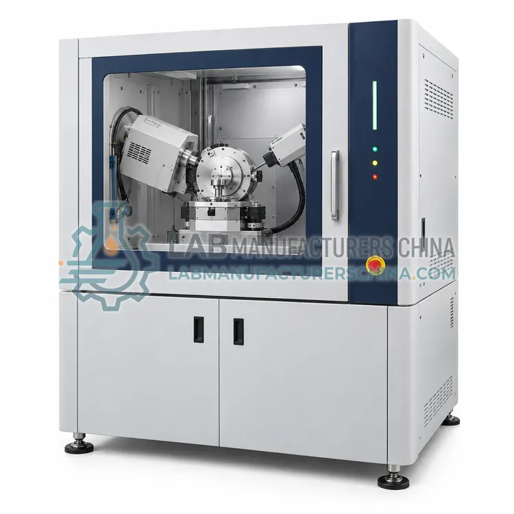
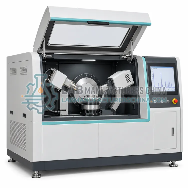
An in-situ X-ray diffractometer adds a controlled sample environment so structural changes can be followed in real time. It is chosen for phase transitions, dehydration, sintering, catalysis, reaction and battery cycling studies.
What is the In-Situ X-Ray Diffractometer with Temperature Stage?
An in-situ X-ray diffractometer adds a controlled sample environment so structural changes can be followed in real time. It is chosen for phase transitions, dehydration, sintering, catalysis, reaction and battery cycling studies.
Overview
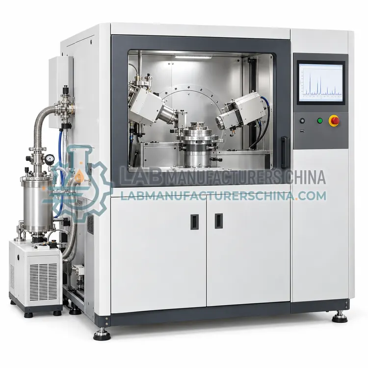
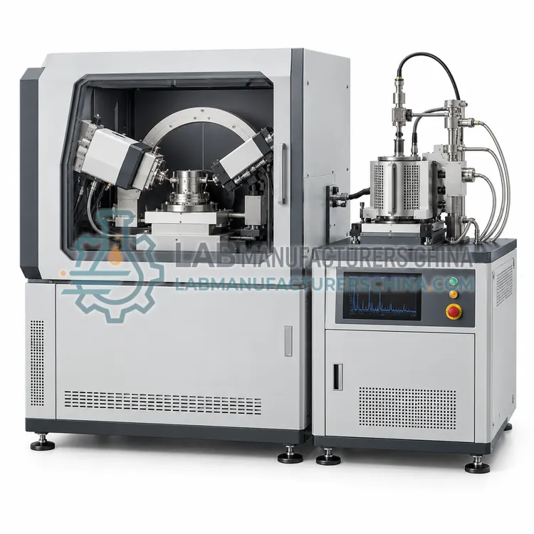
This system couples a diffractometer with a stage that controls temperature, atmosphere or humidity while diffraction patterns are recorded repeatedly. Heating stages reach from ambient to over 1500 degrees Celsius, cooling stages use liquid nitrogen or Peltier elements, and gas or humidity cells simulate reaction or storage conditions. A fast detector and short scans capture time-resolved series, and software aligns and compares patterns to identify phase transitions, reaction pathways, thermal expansion and crystallization. Chamber windows must be transparent to X-rays and mechanically stable, and temperature calibration with a standard is essential. The technique is central to catalyst development, battery electrode studies, cement hydration, metal oxidation and pharmaceutical stability, because it reveals intermediate phases that quenched samples miss.
How does it work?
A focused X-ray beam, usually copper K-alpha radiation, strikes a powdered or polycrystalline sample. Crystal lattice planes scatter the beam, and constructive interference occurs at angles that satisfy Bragg's law, n times lambda equals two d sine theta. The detector records peak positions and intensities as the angle between the beam and the sample changes, producing a diffractogram. Peak positions reveal the lattice spacing and identify phases from reference databases, while peak intensities give relative phase amounts and peak widths relate to crystallite size and microstrain. Preferred orientation, sample displacement and fluorescence can distort results, so sample preparation and instrument alignment are critical. Modern instruments use theta-theta geometry, solid-state or position-sensitive detectors, and software for search-match, Rietveld refinement and profile analysis.
What are the key features?
- Heating stage from ambient to over 1500 °C
- Cooling with liquid nitrogen or Peltier
- Gas, vacuum and humidity cell options
- Fast detector for time-resolved scans
- Software for phase transition analysis
- X-ray transparent chamber windows
What are the technical specifications?
A typical in-situ x-ray diffractometer with temperature stage configuration offers temperature range of -190 to 1600 °C, heating rate of 0.1–100 °C/min, atmospheres of air, inert, vacuum, reactive gas, 2-theta range of 5–120° typical, detector of position-sensitive, fast and scan time of 10 s to 5 min. Values are indicative of the product class; the exact figures for each configuration are confirmed on the quotation and the calibration documents supplied with the instrument.
Which applications is it used for?
The In-Situ X-Ray Diffractometer with Temperature Stage is used in catalyst phase evolution during reaction, battery electrode cycling studies, cement hydration and dehydration and metal oxidation and sintering behavior. Across the wider XRD class, laboratories also deploy it for pharmaceutical polymorph and crystallinity testing, cement, clinker and mineral phase analysis and catalyst, battery and ceramic materials.
Which standards and certifications apply?
Relevant standards and methods include USP <941> X-Ray Diffraction, ASTM E915 Verifying the alignment of X-ray diffraction instrumentation, ASTM D934 Identification of crystalline compounds in water-formed deposits, ISO 20203 Carbonaceous materials for aluminium production — XRD of calcined coke, ISO/IEC 17025 and IEC 61010-2-091. Certification candidates for this model are CE, Radiation safety enclosure and Pressure vessel conformance; each is to be confirmed for the specific configuration before shipment, and certificates are supplied only where they have been issued.
X-ray diffraction instruments are regulated radiation devices and require shielding, interlocks, radiation surveys and trained operators. Powder samples must be ground finely and packed reproducibly, because particle size, packing density and preferred orientation bias peak intensities; back-loading, side-loading or spinning stages reduce orientation effects. Sample fluorescence from iron, cobalt or manganese samples raises background with copper radiation, which may require an alternative wavelength or a monochromator. Alignment, zero-offset and instrument profile are verified with standards such as silicon or corundum. Detectors and X-ray tubes are wear items, and goniometer calibration should be checked periodically. Keep sample preparation records for method traceability.
How do I select the right In-Situ X-Ray Diffractometer with Temperature Stage?
Select an in-situ diffractometer when structural changes with temperature, atmosphere or time are key to the study, and quenched or ex-situ samples would miss intermediate phases. Specify the temperature and atmosphere range, scan speed and window material. Confirm temperature calibration and that the stage fits the diffractometer geometry.
How does it compare with other XRD variants?
Select a powder diffractometer by resolution, detector type, sample environment and throughput. Benchtop instruments are compact and easy to use for phase identification and routine QC but have lower resolution and fewer accessories. High-resolution research systems use long arms, monochromators and high-precision goniometers for accurate lattice parameters, quantitative analysis and complex profile fitting. In-situ systems add heating, cooling, humidity or gas stages for time-resolved studies. Micro-focus instruments provide small beams for inclusions and small samples. Transmission geometry suits weakly absorbing organic and pharmaceutical materials and can reduce preferred orientation. Confirm the 2-theta range, angular resolution, sample changer capacity and software features such as Rietveld and PDF database access for your application.
Full specifications
| Temperature range | -190 to 1600 °C |
|---|---|
| Heating rate | 0.1–100 °C/min |
| Atmospheres | air, inert, vacuum, reactive gas |
| 2-theta range | 5–120° typical |
| Detector | position-sensitive, fast |
| Scan time | 10 s to 5 min |
| Humidity control | optional 5–95% RH |
| Window material | beryllium or kapton |
| Temperature accuracy | ±1 °C with calibration |
| Interface | Ethernet |
Indicative values for this product class. Final specifications are confirmed on the quotation and in the calibration documents supplied with the instrument.
Applications
- Catalyst phase evolution during reaction
- Battery electrode cycling studies
- Cement hydration and dehydration
- Metal oxidation and sintering behavior
Key features
- Heating stage from ambient to over 1500 °C
- Cooling with liquid nitrogen or Peltier
- Gas, vacuum and humidity cell options
- Fast detector for time-resolved scans
- Software for phase transition analysis
- X-ray transparent chamber windows
Frequently asked questions about In-Situ X-Ray Diffractometer with Temperature Stage
Quenching can preserve a metastable phase, cause a phase to transform on cooling, or miss transient intermediates entirely. In-situ diffraction records the sequence of phases at the actual conditions, which is essential for understanding reaction pathways, kinetics and the effect of temperature and atmosphere.
The stage is calibrated with materials of known phase transition or melting temperatures, such as specific salts, and the measured transition is compared with the reference value. Calibration is repeated after window changes or thermocouple replacement, and uncertainty is reported with the data.
With a position-sensitive detector, a usable pattern over a limited 2-theta range can be collected in tens of seconds, allowing time-resolved studies of rapid reactions. Full-range high-resolution scans take longer. The balance between range, resolution and time is chosen for each experiment.
Grind the sample to a fine, uniform powder, then pack it into a holder by back-loading or side-loading so the surface is flat and level with the holder edge. Spinning or using a zero-background holder reduces preferred orientation and background. Preparation quality strongly affects relative peak intensities.
Plate-like or needle-like crystallites align when packed, so some lattice planes diffract more strongly than they should, distorting quantitative results. Gentle grinding, side-loading, spray drying and sample spinning reduce the effect. Rietveld refinement can model and correct residual orientation when the degree is known.
Quantitative phase analysis compares measured peak intensities with reference intensity ratios or applies full-pattern Rietveld refinement using known crystal structures. Rietveld is preferred for complex mixtures because it fits the whole pattern and accounts for overlap, background and preferred orientation. Accuracy depends on good sample preparation and structural models.