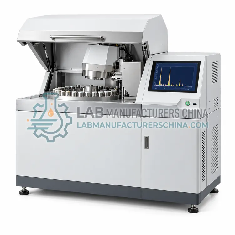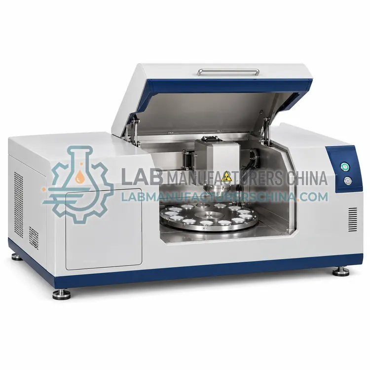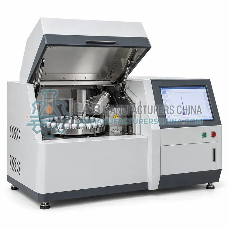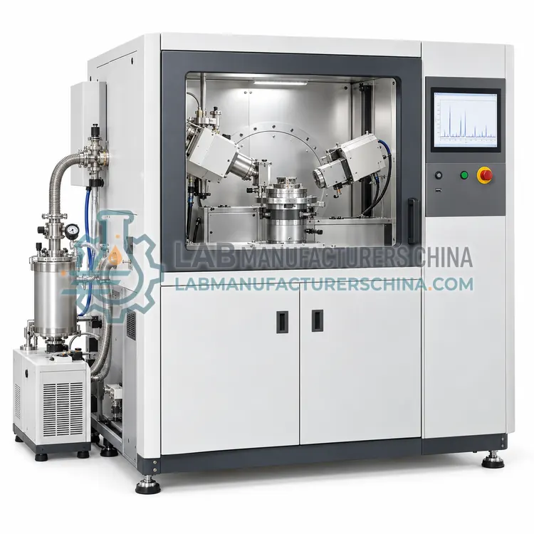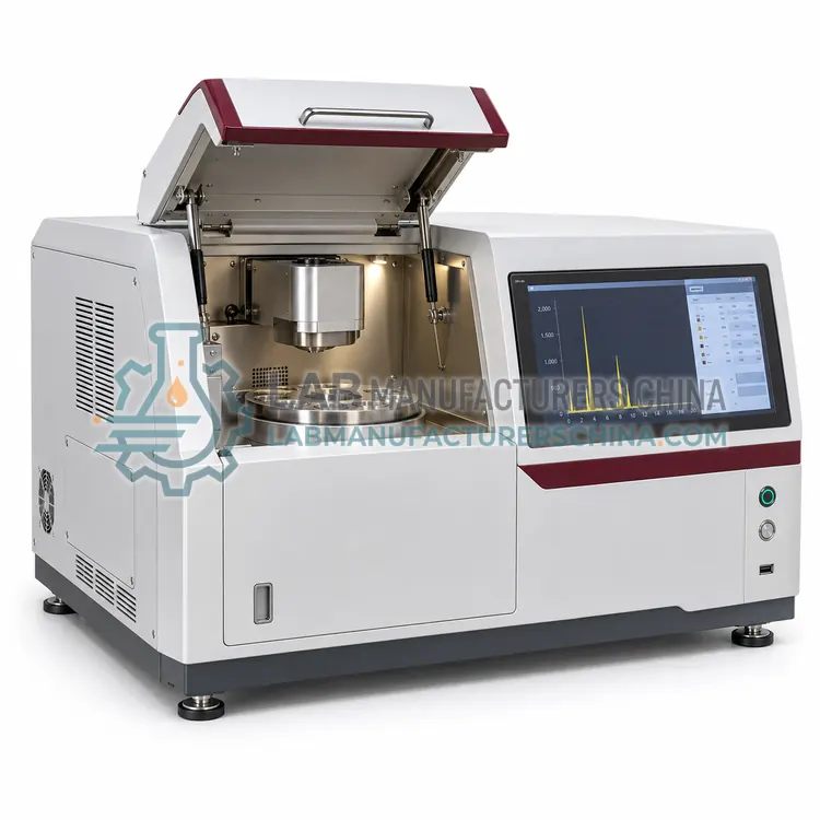
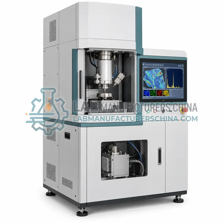
Micro X-Ray Fluorescence Spectrometer
微区 X 射线荧光光谱仪
- SKU XRF-0004
- XRF
- CE (to be confirmed)
- Radiation safety enclosure (to be confirmed)
- ISO 9001 manufacturing (to be confirmed)
Price on request — FOB / CIF quoted per order
Prices depend on configuration, quantity and destination port.
Key specifications
| Spot size | 10–100 µm |
|---|---|
| Element range | Na to U |
| Detection limit | 10–1000 mg/kg typical |
| Stage travel | up to 100 × 100 mm |
| Stage resolution | 1 µm |
| X-ray tube power | 30–100 W |
Request a quote Download brochure · to be provided
By Lab Manufacturers China Technical TeamLast reviewed Standards: ASTM E1621 Wavelength dispersive X-ray fluorescence, ASTM D4294 Sulfur in petroleum products by EDXRF, ISO 3497 Metallic coatings — X-ray spectrometric thickness
What is the Micro X-Ray Fluorescence Spectrometer?
A micro-XRF spectrometer focuses the X-ray beam to a small spot for spatially resolved elemental analysis. It is chosen for microscopic inclusions, coatings, multilayers, forensic traces and heterogeneous samples in materials research.
What is the Micro X-Ray Fluorescence Spectrometer?
A micro-XRF spectrometer focuses the X-ray beam to a small spot for spatially resolved elemental analysis. It is chosen for microscopic inclusions, coatings, multilayers, forensic traces and heterogeneous samples in materials research.
Overview
Polycapillary optics or a similar focusing element concentrates X-rays from the tube onto a spot typically ranging from 10 to 100 micrometres, and the sample is moved under the beam by a motorized stage. Spectra collected point by point build elemental maps across a surface, and line scans reveal coating layering and diffusion profiles. Because the beam is small, the technique can analyze single inclusions in minerals, contamination particles, solder joints, art and archaeological objects, and forensic trace evidence without damaging them. Element sensitivity is lower than conventional XRF because the beam is concentrated, so measurement times are longer. Vacuum or helium purge is used for light elements, and stage reproducibility determines the spatial accuracy of maps.
How does it work?
A primary X-ray beam from a tube or radioisotope source ejects inner-shell electrons from sample atoms. Electrons from higher shells fall into the vacancies and emit X-ray photons whose energies equal the difference between the two levels, which is unique to each element. The intensity of each characteristic line is proportional to the amount of that element, so calibration with reference materials gives concentration. Energy-dispersive instruments use a semiconductor detector such as silicon drift to measure photon energies directly and record the whole spectrum at once. Wavelength-dispersive instruments use analyzing crystals to diffract specific wavelengths to a detector, giving better resolution and lower detection limits, especially for light elements. Matrix effects from absorption and secondary excitation are corrected by fundamental parameters or empirical influence coefficients, and sample form is matched to calibration standards.
What are the key features?
- Polycapillary focusing optics with small spot
- Motorized stage for point, line and map scans
- Elemental mapping of heterogeneous samples
- Coating and multilayer thickness profiling
- Non-destructive analysis of inclusions and particles
- Vacuum or helium purge for light elements
What are the technical specifications?
A typical micro x-ray fluorescence spectrometer configuration offers spot size of 10–100 µm, element range of Na to U, detection limit of 10–1000 mg/kg typical, stage travel of up to 100 × 100 mm, stage resolution of 1 µm and X-ray tube power of 30–100 W. Values are indicative of the product class; the exact figures for each configuration are confirmed on the quotation and the calibration documents supplied with the instrument.
Which applications is it used for?
The Micro X-Ray Fluorescence Spectrometer is used in mineral inclusions and geological phases, coating, solder and multilayer analysis, forensic trace evidence and contamination and art, archaeology and cultural heritage objects. Across the wider XRF class, laboratories also deploy it for elemental analysis of alloys, ceramics and glass, roHS and restricted substance screening and coating thickness and thin-film composition.
Which standards and certifications apply?
Relevant standards and methods include ASTM E1621 Wavelength dispersive X-ray fluorescence, ASTM D4294 Sulfur in petroleum products by EDXRF, ISO 3497 Metallic coatings — X-ray spectrometric thickness, IEC 62321 Determination of restricted substances in electrotechnical products, US EPA Method 6200 Field portable XRF for soil, ISO 13196 Soil quality — Screening of selected elements by EDXRF, IEC 61010-2-091 and ISO/IEC 17025. Certification candidates for this model are CE, Radiation safety enclosure and ISO 9001 manufacturing; each is to be confirmed for the specific configuration before shipment, and certificates are supplied only where they have been issued.
XRF instruments are regulated radiation devices, so installation requires shielding, interlocks, radiation surveys and trained operators, with portable units also needing safe work procedures. Sample preparation strongly affects accuracy: surface contamination, insufficient thickness and particle size effects bias results, and powders are best pressed or fused. Calibration must match the sample matrix, and drift is monitored with check standards. Vacuum or helium systems need clean seals and gas supplies for light elements. Detector windows and X-ray tubes are wear parts with finite life. Keep records of tube hours, calibration and checks, and dispose of X-ray components according to radiation regulations.
How do I select the right Micro X-Ray Fluorescence Spectrometer?
Select micro-XRF when elemental information is needed at the microscopic scale, for example inclusions, particles, coatings or layered structures, and the sample must remain intact. Specify the spot size, map area and element range. Confirm stage reproducibility and that light-element sensitivity meets your needs, which may require vacuum or helium.
How does it compare with other XRF variants?
Select an XRF configuration by element range, detection limit and sample type. Energy-dispersive instruments are compact, fast and well suited to routine screening and multi-element analysis, with moderate resolution. Wavelength-dispersive instruments cost more and are slower but excel for light elements, precise major-oxide analysis and low detection limits. Total reflection instruments reach ultratrace levels on small sample spots and serve semiconductor, water and biological analysis. Polarized energy-dispersive instruments lower background for trace work without the cost of wavelength dispersive optics. Micro-XRF adds spatial resolution for inclusions, coatings and forensic samples. Consider sample size, whether vacuum or helium purge is needed for light elements, automation level and the regulatory method your laboratory must follow before deciding.
Full specifications
| Spot size | 10–100 µm |
|---|---|
| Element range | Na to U |
| Detection limit | 10–1000 mg/kg typical |
| Stage travel | up to 100 × 100 mm |
| Stage resolution | 1 µm |
| X-ray tube power | 30–100 W |
| Detector | silicon drift detector |
| Mapping | point, line and area maps |
| Atmosphere | air, vacuum or helium |
| Interface | Ethernet |
Indicative values for this product class. Final specifications are confirmed on the quotation and in the calibration documents supplied with the instrument.
Applications
- Mineral inclusions and geological phases
- Coating, solder and multilayer analysis
- Forensic trace evidence and contamination
- Art, archaeology and cultural heritage objects
Key features
- Polycapillary focusing optics with small spot
- Motorized stage for point, line and map scans
- Elemental mapping of heterogeneous samples
- Coating and multilayer thickness profiling
- Non-destructive analysis of inclusions and particles
- Vacuum or helium purge for light elements
Frequently asked questions about Micro X-Ray Fluorescence Spectrometer
Micro-XRF has lower spatial resolution, typically 10 to 100 micrometres against nanometres for electron microscopy, but it works at atmospheric pressure, needs no conductive coating and penetrates deeper. It is better for hydrated or insulating samples and for below-surface analysis, while electron methods excel for very fine features.
Focusing the beam onto a small spot reduces the total number of photons reaching the sample and increases local beam density, so fewer counts are collected and measurement times must increase. Some elements also show altered excitation efficiency. Sensitivity is therefore lower than conventional XRF with large spots.
Yes, line scans across a polished cross-section or through a coating reveal the intensity profile of coating and substrate elements, from which layer thickness can be estimated with calibration. It is often used alongside dedicated coating thickness gauges for solder, plating and multilayer films.
EDXRF measures photon energies directly with a semiconductor detector and records the whole spectrum simultaneously, giving speed and a compact design. WDXRF uses analyzing crystals to diffract selected wavelengths and offers higher resolution, better light-element performance and lower detection limits at higher cost and longer measurement times.
The sample must be thick enough that the full primary beam is absorbed within it and fluorescence from deeper layers does not change the result, a condition called infinite thickness. This thickness depends on the element and matrix, ranging from micrometres for heavy elements in light matrices to millimetres for light elements, and it is verified during method development.
The intensity of an element line depends not only on its concentration but also on how other elements absorb the primary and fluorescent X-rays and on secondary excitation. These matrix effects can change intensities by large factors, so accurate analysis requires matrix-matched standards or correction models based on fundamental parameters or influence coefficients.
Related models