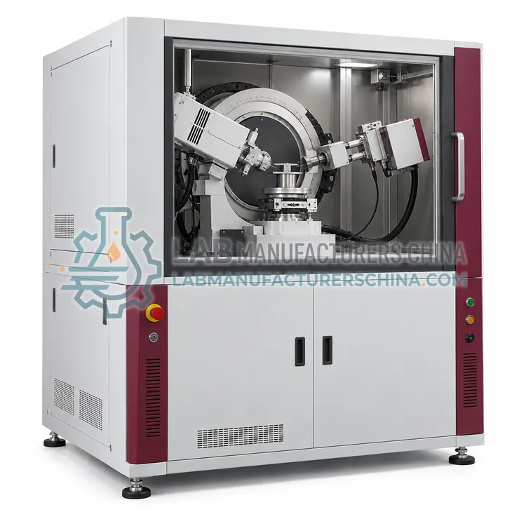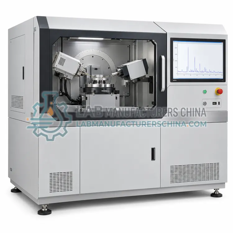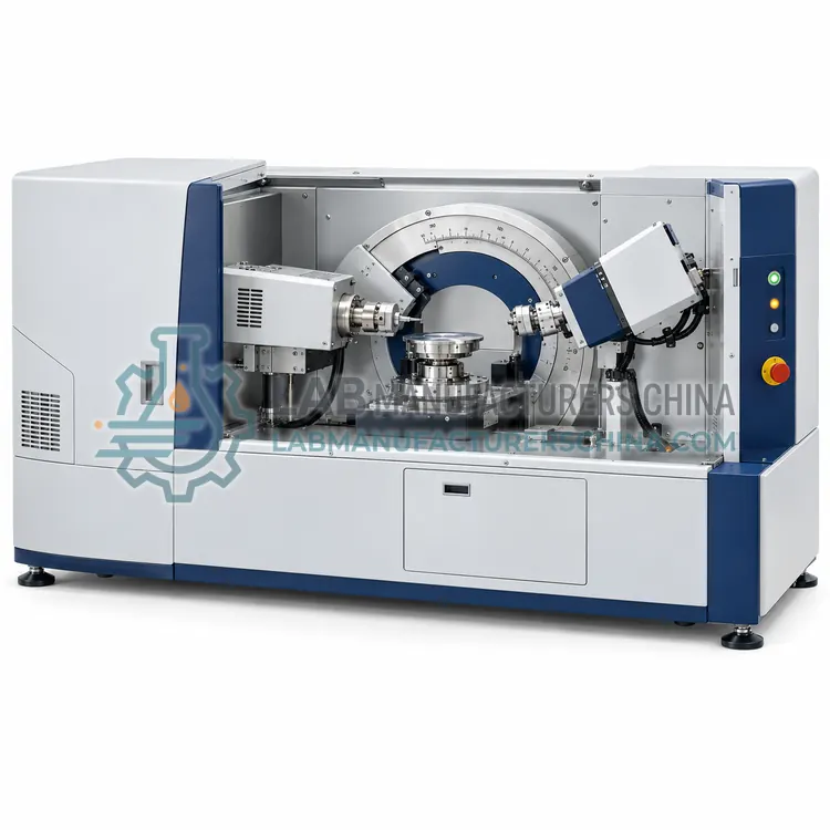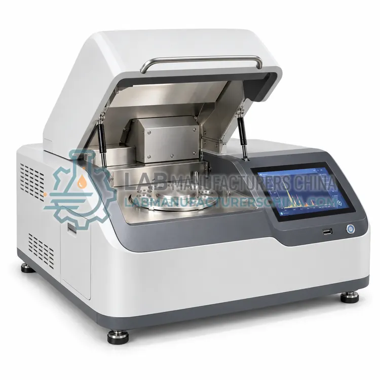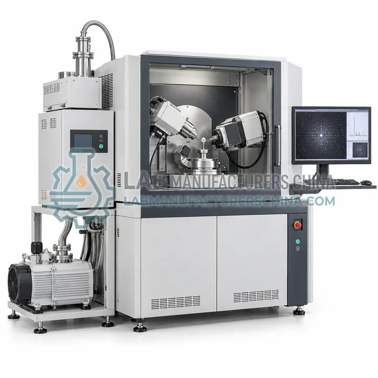
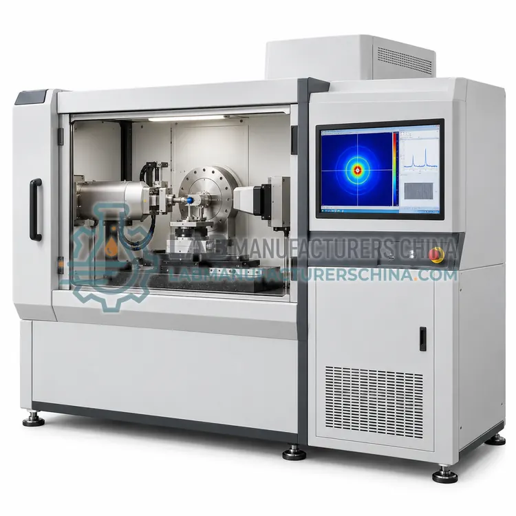
Small-Angle X-Ray Scattering Diffractometer
小角 X 射线散射仪
- SKU XRA-0006
- X-Ray Diffractometers
- CE (to be confirmed)
- Radiation safety enclosure (to be confirmed)
- ISO 9001 manufacturing (to be confirmed)
Price on request — FOB / CIF quoted per order
Prices depend on configuration, quantity and destination port.
Key specifications
| q range | 0.01–5 nm-1 |
|---|---|
| Size range | 1–100 nm |
| Camera length | 0.3–3 m |
| Detector | two-dimensional area detector |
| X-ray source | microfocus tube or synchrotron |
| Beam size | 0.2–1.0 mm |
Request a quote Download brochure · to be provided
By Lab Manufacturers China Technical TeamLast reviewed Standards: ASTM E915 Verifying the alignment of X-ray diffraction instrumentation, ASTM E1426 Determining the effective elastic parameter for X-ray diffraction measurements of residual stress, USP <941> X-Ray Diffraction
What is the Small-Angle X-Ray Scattering Diffractometer?
A small-angle X-ray scattering diffractometer measures scattering at very low angles to resolve nanoscale structures. It is chosen for routine particle size, pores, lamellae and protein shape in solutions, polymers, colloids and nanomaterials.
What is the Small-Angle X-Ray Scattering Diffractometer?
A small-angle X-ray scattering diffractometer measures scattering at very low angles to resolve nanoscale structures. It is chosen for routine particle size, pores, lamellae and protein shape in solutions, polymers, colloids and nanomaterials.
Overview
Small-angle scattering uses a finely collimated beam and a long, usually evacuated, camera to reach angles of a fraction of a degree. Features from about one to one hundred nanometres scatter in this region, so particle size and shape, pore structure, lamellar spacing, aggregation and the overall size of proteins in solution can be determined without crystals. A two-dimensional detector records isotropic or anisotropic patterns, and software fits models or transforms data to size distributions. A beam stop protects the detector from the direct beam, and background subtraction of solvent or matrix is essential. Instruments range from compact laboratory units to synchrotron beamlines, and the technique is central to polymer, nanoparticle, pharmaceutical and structural biology research.
How does it work?
X-rays scatter from the electron density of a material. In a crystal, scattering from periodic lattice planes interferes constructively at angles given by Bragg's law, producing diffraction peaks. A diffractometer positions the sample and detector with high angular precision and records intensity versus angle or versus a two-dimensional detector image. Single-crystal instruments determine atomic structure from the full set of reflections, while powder and thin-film instruments analyze phase mixtures, preferred orientation and layer structure. Peak positions give lattice parameters and strain, peak widths relate to crystallite size and microstrain, and low-angle scattering reveals nanoscale features. The choice of optics, monochromator, detector and sample stage determines which measurements are possible, and instrument alignment with certified standards underpins accuracy.
What are the key features?
- Long evacuated camera for very low angles
- Two-dimensional detector for isotropic and anisotropic patterns
- Size range from about 1 to 100 nm
- Model fitting and size distribution software
- Beam stop and background subtraction
- Compact laboratory or synchrotron configuration
What are the technical specifications?
A typical small-angle x-ray scattering diffractometer configuration offers q range of 0.01–5 nm-1, size range of 1–100 nm, camera length of 0.3–3 m, detector of two-dimensional area detector, X-ray source of microfocus tube or synchrotron and beam size of 0.2–1.0 mm. Values are indicative of the product class; the exact figures for each configuration are confirmed on the quotation and the calibration documents supplied with the instrument.
Which applications is it used for?
The Small-Angle X-Ray Scattering Diffractometer is used in nanoparticle and colloid size and shape, protein and macromolecule solution structure, polymer and block copolymer morphology and porous materials and lamellar structures. Across the wider X-Ray Diffractometers class, laboratories also deploy it for single-crystal structure determination, thin-film thickness, texture and phase analysis and residual stress in welds, coatings and machined parts.
Which standards and certifications apply?
Relevant standards and methods include ASTM E915 Verifying the alignment of X-ray diffraction instrumentation, ASTM E1426 Determining the effective elastic parameter for X-ray diffraction measurements of residual stress, USP <941> X-Ray Diffraction, ISO 20203 Carbonaceous materials — XRD of calcined coke, ISO/IEC 17025 and IEC 61010-2-091. Certification candidates for this model are CE, Radiation safety enclosure and ISO 9001 manufacturing; each is to be confirmed for the specific configuration before shipment, and certificates are supplied only where they have been issued.
These are regulated radiation devices requiring shielding, interlocks, radiation surveys and trained operators. Single crystals must be mounted and centered precisely, and low-temperature or humidity devices often protect them. Thin films and wafers need flat mounting and careful alignment to the beam, and reflectometry requires very low background and precise angular steps. Residual stress methods need defined measurement directions and elastic constants. Small-angle systems require long evacuated paths and clean beam stops. Alignment, angular calibration and detector linearity are verified with standards, and X-ray tubes, goniometer motors and area detectors are wear items. Sample preparation and measurement conditions should be recorded for traceability.
How do I select the right Small-Angle X-Ray Scattering Diffractometer?
Select a small-angle scattering instrument when nanometre-scale size, shape or ordering must be measured in solutions, polymers or powders without crystallizing the sample. Specify the size range, sample form and throughput. For very weak scatterers or the smallest angles, consider synchrotron access; for routine work, a laboratory instrument is usually sufficient.
How does it compare with other X-Ray Diffractometers variants?
Select a diffractometer configuration by the structural question. Single-crystal systems with four-circle or kappa goniometers and area detectors determine atomic structures from small crystals. Thin-film instruments with grazing-incidence optics and parallel beams measure layer thickness, texture and phase in coatings. Reflectometry configurations measure electron density profiles, interface roughness and layer thickness at very low angles. Residual stress instruments use side-inclination or psi geometry with precise sample tilting to measure strain in metals and ceramics. Small-angle scattering systems with long camera lengths resolve nanometre-scale particles, pores and lamellae. Confirm the angular range and resolution, sample environment, detector type and software for your application, and verify that the instrument can follow any required standard method.
Full specifications
| q range | 0.01–5 nm-1 |
|---|---|
| Size range | 1–100 nm |
| Camera length | 0.3–3 m |
| Detector | two-dimensional area detector |
| X-ray source | microfocus tube or synchrotron |
| Beam size | 0.2–1.0 mm |
| Sample types | solutions, polymers, powders, films |
| Temperature control | optional 5–60 °C stage |
| Measurements per hour | 5–60 |
| Interface | Ethernet |
Indicative values for this product class. Final specifications are confirmed on the quotation and in the calibration documents supplied with the instrument.
Applications
- Nanoparticle and colloid size and shape
- Protein and macromolecule solution structure
- Polymer and block copolymer morphology
- Porous materials and lamellar structures
Key features
- Long evacuated camera for very low angles
- Two-dimensional detector for isotropic and anisotropic patterns
- Size range from about 1 to 100 nm
- Model fitting and size distribution software
- Beam stop and background subtraction
- Compact laboratory or synchrotron configuration
Frequently asked questions about Small-Angle X-Ray Scattering Diffractometer
Small-angle scattering resolves features from roughly one to one hundred nanometres, bridging the gap between crystallography and light scattering. The accessible range depends on the q range, which is set by beam energy and camera length. Larger structures need ultra-small-angle configurations.
The scattering from solvent, buffer or matrix is often comparable to or larger than that from the particles of interest, especially for dilute solutions. Measuring the same solvent separately and subtracting it isolates the particle signal. Cell windows, air and beam radiation damage also contribute background.
Both use X-ray scattering, but XRD measures sharp diffraction peaks from periodic crystal planes at wider angles, while SAXS measures diffuse or ordered scattering at very low angles from structures much larger than atomic spacing. SAXS does not require crystals and is used for particles, pores and polymers.
Single-crystal diffraction measures individual reflections from one crystal, giving enough data to solve the full atomic structure, including bond lengths and angles. Powder diffraction measures the overlapping reflections from many randomly oriented crystallites, which identifies and quantifies phases but cannot alone determine a complete atomic structure.
The sample is tilted to several angles and the shift of a chosen diffraction peak is measured at each tilt. Lattice strain is calculated from the peak shift, and stress follows from the strain and the material elastic constants. Methods follow standards such as ASTM E1426 and require defined measurement directions.
At very shallow incidence angles the X-ray beam probes only the near-surface region of a thin film or coating, which reduces signal from the substrate. This allows phase identification, texture and depth profiling of layers only nanometres to micrometres thick, as used for semiconductors, optical coatings and corrosion films.