Western Blot Chemiluminescence Imaging System
Western blot 化学发光成像系统
- SKU PRA-0004
- Protein Analysis
- CE (to be confirmed)
- ISO 9001 manufacturing (to be confirmed)
Price on request — FOB / CIF quoted per order
Prices depend on configuration, quantity and destination port.
Key specifications
| Camera sensor | Cooled CCD/CMOS, 4–9 MP |
|---|---|
| Cooling | −25 to −60 °C below ambient |
| Dynamic range | 16-bit, ≥4 orders |
| Exposure time | 0.001 s–30 min |
| Imaging area | up to 20 × 24 cm |
| Detection modes | Chemiluminescence; fluorescence optional |
Request a quote Download brochure · to be provided
By Lab Manufacturers China Technical TeamLast reviewed Standards: IEC 61010-1, IEC 61010-2-101, IEC 61326-2-6
What is the Western Blot Chemiluminescence Imaging System?
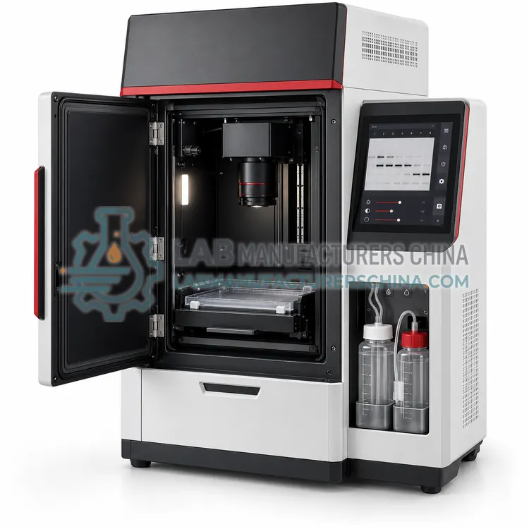
A Western blot chemiluminescence imaging system uses a cooled CCD or CMOS camera to capture chemiluminescent and fluorescent signals from membranes with exposures from seconds to several minutes. It replaces X-ray film for protein detection and supports quantitative densitometry within the linear range.
What is the Western Blot Chemiluminescence Imaging System?
A Western blot chemiluminescence imaging system uses a cooled CCD or CMOS camera to capture chemiluminescent and fluorescent signals from membranes with exposures from seconds to several minutes. It replaces X-ray film for protein detection and supports quantitative densitometry within the linear range.
Overview
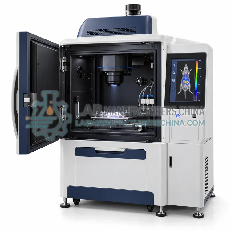
The imaging system places a membrane in a light-tight chamber under a high-aperture lens coupled to a cooled camera, typically cooled to −25 to −60 °C below ambient to suppress dark noise. Chemiluminescent substrates, such as enhanced luminol, emit light for several minutes after HRP reaction, which the camera integrates during long exposure. Multichannel models add LED or laser excitation and emission filters for fluorescent Western blots and stain-free total protein normalization. Dynamic range of 4 or more orders allows quantification without saturation, and automatic exposure sequences capture multiple images for selecting unsaturated signals. Software provides band detection, lane profiling, molecular weight estimation and normalization to loading controls. Data can be exported as TIFF files suitable for publication with raw image retention.
How does it work?
Protein analysis relies on several complementary physical principles. Microplate readers measure absorbance, fluorescence or luminescence from wells to quantify protein by colorimetric assays such as BCA and Bradford or to read ELISA signals, using monochromators or filters and photomultiplier tubes or photodiodes. Electrophoretic systems separate proteins by size, charge or isoelectric point; in capillary systems, SDS-coated proteins migrate through a sieving polymer under high voltage and are detected by UV absorbance or laser-induced fluorescence. Western blot imagers capture chemiluminescent or fluorescent signals from membranes using cooled CCD cameras with long exposure. Chromatography systems such as FPLC pump buffers through columns to bind and elute proteins by affinity, ion exchange or size exclusion, with UV, conductivity and pH monitors guiding fraction collection.
What are the key features?
- Cooled camera suppresses noise for long chemiluminescence exposures.
- Dynamic range of four or more orders supports quantification without saturation.
- Automatic exposure sequences capture unsaturated signals.
- Optional fluorescence channels enable multiplex blots and total protein normalization.
- Software for lane profiling, band quantification and molecular weight estimation.
- Raw data retention with TIFF export for publication.
What are the technical specifications?
A typical western blot chemiluminescence imaging system configuration offers camera sensor of Cooled CCD/CMOS, 4–9 MP, cooling of −25 to −60 °C below ambient, dynamic range of 16-bit, ≥4 orders, exposure time of 0.001 s–30 min, imaging area of up to 20 × 24 cm and detection modes of Chemiluminescence; fluorescence optional. Values are indicative of the product class; the exact figures for each configuration are confirmed on the quotation and the calibration documents supplied with the instrument.
Which applications is it used for?
The Western Blot Chemiluminescence Imaging System is used in chemiluminescent Western blot detection, multiplex fluorescent Western blotting, total protein normalization and dot blot and colony imaging. Across the wider Protein Analysis class, laboratories also deploy it for ELISA and immunoassay readout, protein concentration determination and antibody purity and size variant analysis.
Which standards and certifications apply?
Relevant standards and methods include IEC 61010-1, IEC 61010-2-101, IEC 61326-2-6, USP <1053>, USP <1055>, Ph. Eur. 2.2.31 and ICH Q6B. Certification candidates for this model are CE and ISO 9001 manufacturing; each is to be confirmed for the specific configuration before shipment, and certificates are supplied only where they have been issued.
Protein instruments depend on good sample handling: keep samples cold, use protease inhibitors and avoid repeated freeze-thaw cycles. Microplate readers need periodic verification of wavelength accuracy and absorbance linearity with reference filters or dye solutions. Capillary systems require clean buffers, filtered samples and replacement of cartridges on schedule. Imaging systems should be kept in a dust-free, temperature-stable location, and cameras need flat-field correction. FPLC columns must be stored in recommended solutions, and pump seals replaced regularly. Regulated laboratories should perform installation and operational qualification, and consider 21 CFR Part 11 compliant software.
How do I select the right Western Blot Chemiluminescence Imaging System?
Choose a Western blot imager when film is being replaced or quantitative densitometry is required. Specify camera cooling, dynamic range, imaging area, fluorescence channels and software normalization features. For nucleic acid gel imaging only, a gel documentation system is sufficient.
How does it compare with other Protein Analysis variants?
Define the analytical goal first: quantification and ELISA point to microplate readers, purity and size variants to capillary electrophoresis, expression and semi-quantitative detection to Western blot imaging, and isolation of milligram quantities to chromatography systems. Check sensitivity and dynamic range against your lowest expected signal, for example pg/mL for ELISA or ng per band for blotting. Consider throughput and automation, such as 96-sample capillary runs or plate stackers. For purification, specify column pressure limits, flow rate range and detector options. Confirm compatibility with your assay formats and reagents, and evaluate software for data integrity, regulatory audit trail and export. Factor in consumable costs, such as capillary cartridges or detection reagents, and available training.
Full specifications
| Camera sensor | Cooled CCD/CMOS, 4–9 MP |
|---|---|
| Cooling | −25 to −60 °C below ambient |
| Dynamic range | 16-bit, ≥4 orders |
| Exposure time | 0.001 s–30 min |
| Imaging area | up to 20 × 24 cm |
| Detection modes | Chemiluminescence; fluorescence optional |
| Fluorescence channels | 2–4 (optional) |
| Lens | f/0.95–f/1.2, motorized zoom and focus |
| Data format | 16-bit TIFF |
| Power supply | 100–240 V, 50/60 Hz |
Indicative values for this product class. Final specifications are confirmed on the quotation and in the calibration documents supplied with the instrument.
Applications
- Chemiluminescent Western blot detection
- Multiplex fluorescent Western blotting
- Total protein normalization
- Dot blot and colony imaging
Key features
- Cooled camera suppresses noise for long chemiluminescence exposures.
- Dynamic range of four or more orders supports quantification without saturation.
- Automatic exposure sequences capture unsaturated signals.
- Optional fluorescence channels enable multiplex blots and total protein normalization.
- Software for lane profiling, band quantification and molecular weight estimation.
- Raw data retention with TIFF export for publication.
Frequently asked questions about Western Blot Chemiluminescence Imaging System
Cooling reduces thermal noise, allowing long exposures of several minutes without excessive background. This improves detection of weak bands and widens dynamic range, which matters because chemiluminescent signals are faint and time-limited.
Image within the linear range without saturated pixels, normalize to total protein or a validated loading control, run replicates and use consistent exposure and background subtraction. Total protein normalization is increasingly preferred to single housekeeping proteins.
Many systems support gel imaging with epi-white, UV or blue light illumination and appropriate filters, but performance depends on the optics and options. Confirm that the light sources needed for your stains are included before purchase.
Common instruments include microplate readers for protein quantification and ELISA, capillary electrophoresis systems for purity and size variants, Western blot imagers for expression detection and FPLC systems for purification. Choice depends on the information needed, sensitivity and sample number.
BCA relies on copper reduction by protein and is compatible with most detergents but sensitive to reducing agents. Bradford uses Coomassie dye binding, is faster and tolerates reducing agents, but is incompatible with many detergents and varies more between proteins.
CE-SDS is capillary electrophoresis of SDS-denatured proteins in a sieving matrix, separating them by molecular weight. It replaces slab SDS-PAGE in biopharma for quantitative purity and fragment analysis of antibodies, giving reproducible, automated and validated results.
Related models
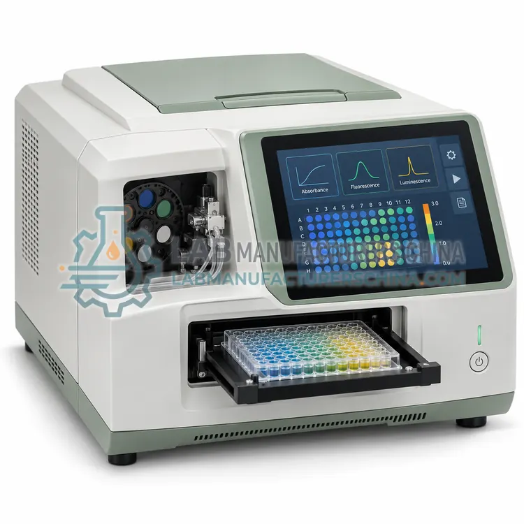
Multimode Microplate Reader (Absorbance, Fluorescence, Luminescence)
多功能酶标仪(吸光度、荧光、发光)
- CE (to be confirmed)
- +2