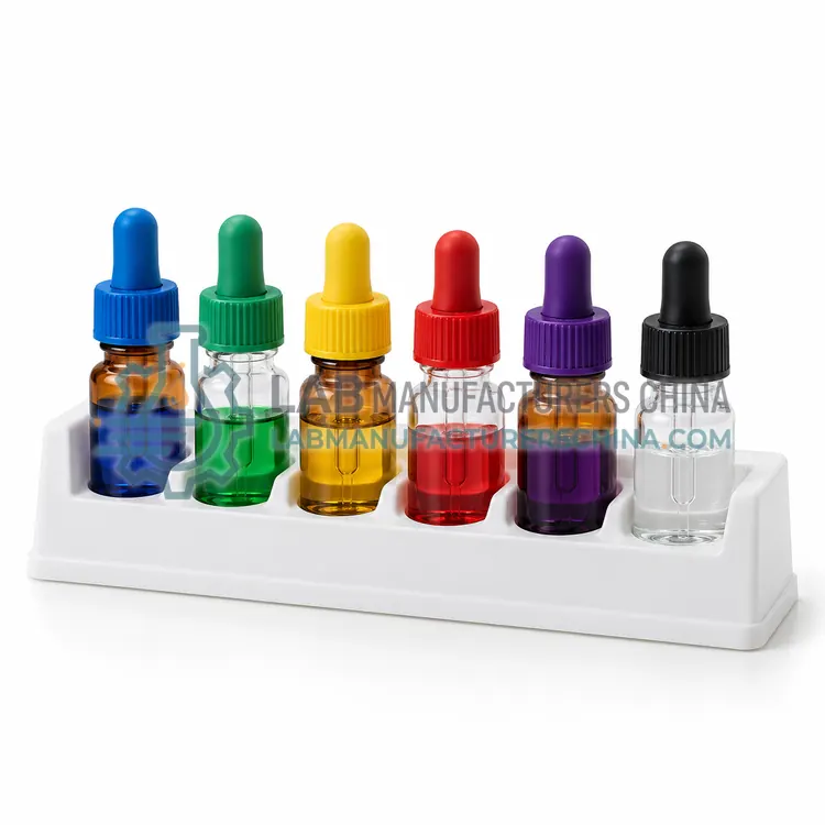
Fluorochrome-Conjugated Monoclonal Antibody Panel Reagents
荧光素偶联单克隆抗体检测组合试剂
- ISO 13485 manufacturing (to be confirmed)
- +2
Welcome to Lab Manufacturers China — laboratory instruments direct from a Chinese manufacturer
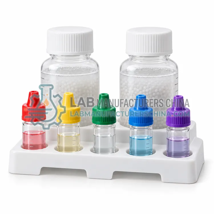
补偿与校准微球试剂
Price on request — FOB / CIF quoted per order
Prices depend on configuration, quantity and destination port.
| Bead material | Polystyrene microspheres |
|---|---|
| Bead diameter | Approx. 3–8 µm |
| Capture reactivity | Mouse, rat, hamster or human Ig (kit dependent) |
| Bead concentration | Approx. 1 × 10^6 beads/mL (compensation); approx. 1,000 beads/µL (counting) |
| Peaks per calibration set | 3–8 peaks |
| CV of bead population | Below 3% (typical) |
By Lab Manufacturers China Technical TeamLast reviewed Standards: CLSI H62, CLSI H43, ISO 20391-2
Compensation and calibration beads are uniform polystyrene microspheres used to set compensation matrices and to monitor cytometer performance. Antibody-capture beads provide bright positive and negative populations for any conjugate, while fluorescent calibration beads verify laser alignment, sensitivity and quantitation.
Compensation and calibration beads are uniform polystyrene microspheres used to set compensation matrices and to monitor cytometer performance. Antibody-capture beads provide bright positive and negative populations for any conjugate, while fluorescent calibration beads verify laser alignment, sensitivity and quantitation.
Compensation beads are polystyrene microspheres, typically 3 to 8 micrometers in diameter, coated with anti-immunoglobulin capture antibodies that bind any conjugated antibody of the matching species, giving one positive and one negative population with consistent brightness. This avoids consuming precious cells for single-stain controls and allows tandem dyes to be compensated accurately. Calibration beads are different: they carry fixed fluorochrome amounts, such as 3 to 6 peak sets, for daily quality control of laser alignment, detector linearity and sensitivity, or MESF beads that map fluorescence to molecules of equivalent soluble fluorochrome. Counting beads of known concentration, supplied at about 1,000 beads per microliter, allow absolute cell counts to be calculated from bead-to-cell event ratios. Beads are supplied in dropper bottles or vials and stored at 2 to 8 degrees, protected from light.
Cytometry reagents make cellular properties detectable as optical signals. Monoclonal antibodies conjugated to fluorochromes such as FITC, PE, APC, or tandem and polymer dyes bind specific antigens, and the laser-excited fluorescence is proportional to antigen density. Nucleic acid and amine-reactive dyes discriminate live from dead cells, because membrane-compromised cells take up DNA dyes or expose more amines. Calibration and compensation beads bind antibodies through capture moieties or carry defined fluorochrome content, providing consistent positive and negative populations for compensation matrices and standardization. Fixation cross-links proteins, often with formaldehyde, while permeabilization with detergents or alcohol allows antibodies to reach intracellular or nuclear targets. Lysing solutions remove red blood cells by osmotic or ammonium-chloride lysis while preserving leukocyte scatter properties.
A typical compensation and calibration bead reagents configuration offers bead material of Polystyrene microspheres, bead diameter of Approx. 3–8 µm, capture reactivity of Mouse, rat, hamster or human Ig (kit dependent), bead concentration of Approx. 1 × 10^6 beads/mL (compensation); approx. 1,000 beads/µL (counting), peaks per calibration set of 3–8 peaks and CV of bead population of Below 3% (typical). Values are indicative of the product class; the exact figures for each configuration are confirmed on the quotation and the calibration documents supplied with the instrument.
The Compensation and Calibration Bead Reagents is used in compensation matrix setup, daily cytometer quality control, absolute cell counting, MESF quantitation of antigen density and instrument standardization across sites. Across the wider Cytometry Reagents class, laboratories also deploy it for immunophenotyping panels, live/dead cell discrimination and instrument compensation and QC.
Relevant standards and methods include CLSI H62, CLSI H43, ISO 20391-2, ISO 13485 (IVD reagent manufacturing), EU IVDR 2017/746 and ISAC MIFlowCyt. Certification candidates for this model are ISO 13485 manufacturing, CE-IVD marking for selected products and ISO 9001 manufacturing; each is to be confirmed for the specific configuration before shipment, and certificates are supplied only where they have been issued.
Store antibodies and dyes at 2 to 8 degrees Celsius, protected from light, and never freeze tandem dyes, which degrade and cause spillover. Titrate every new antibody lot, and record lot numbers in your experiment records. Use Fc receptor blocking where needed, and include fluorescence-minus-one and isotype or biological controls when setting gates. Prepare fresh formaldehyde solutions and validate fixation time for each epitope. Dispose of azide-containing buffers and lysing reagents according to local chemical regulations, as sodium azide can form explosive metal azides in plumbing. For regulated assays, source reagents with certificates of analysis, and revalidate panels when changing vendors or clones.
Choose capture beads for compensation, fixed-peak fluorescent beads for daily QC, MESF beads for quantitation, and counting beads for absolute counts. Bead type must match antibody species and instrument lasers. In an RFQ specify function, species reactivity, number of peaks, bead concentration and pack size.
Choose reagents to match the instrument's lasers and filters, since fluorochromes must be excited and detected by your configuration. Assign bright fluorochromes to low-density antigens and dim ones to abundant antigens, and avoid spectral overlap between tandem dyes. Confirm antibody clone, species reactivity and validated application for flow cytometry. For intracellular or phospho targets, select compatible fixation and permeabilization buffers. Include a viability dye in every panel, and use compatible compensation beads for each fluorochrome. Check lot-to-lot consistency documentation, storage conditions at 2 to 8 degrees Celsius, expiry dating and supply of bulk or lyophilized formats for regulated assays. Titrate each antibody in your own cells to find the optimal staining concentration.
| Bead material | Polystyrene microspheres |
|---|---|
| Bead diameter | Approx. 3–8 µm |
| Capture reactivity | Mouse, rat, hamster or human Ig (kit dependent) |
| Bead concentration | Approx. 1 × 10^6 beads/mL (compensation); approx. 1,000 beads/µL (counting) |
| Peaks per calibration set | 3–8 peaks |
| CV of bead population | Below 3% (typical) |
| Pack size | 50–200 tests or 2–10 mL |
| Excitation range | 355–640 nm |
| Storage temperature | 2–8 °C, protected from light |
| Shelf life | 12–24 months |
Indicative values for this product class. Final specifications are confirmed on the quotation and in the calibration documents supplied with the instrument.
Beads give consistent, bright positive and clean negative populations without consuming precious cells. They also accommodate tandem dyes whose brightness on cells may be too low, but the same antibody-fluorochrome conjugate and acquisition settings must be used as in the experiment.
Counting beads have a known concentration. Adding a fixed volume to a sample lets you compute absolute cells per microliter from the ratio of cell events to bead events, without a volumetric pump, as used in CD4, CD34 and viable cell counting.
Run fixed-peak calibration beads every day of operation, before samples, to verify laser alignment, sensitivity and fluidics stability. Track median fluorescence and CV over time, and investigate drift before it affects data.
A typical experiment needs fluorochrome-conjugated antibodies, a viability dye, staining buffer with protein, and Fc block. Intracellular staining adds fixation and permeabilization buffers, while compensation beads, counting beads and lysing solutions support calibration, absolute counts and whole blood work.
Match each fluorochrome to your instrument's lasers and detectors, pair bright dyes with weakly expressed antigens, and minimize spectral overlap between channels. Use a spillover spreading matrix or panel design tool to check spreading error before finalizing a panel.
Too much antibody increases nonspecific binding and background, while too little reduces the signal and staining index. Titration with serial dilutions on your own cells identifies the concentration with the best separation of positive and negative populations and saves reagent.
荧光素偶联单克隆抗体检测组合试剂
符合 21 CFR Part 11 的临床流式细胞术软件