How do Automated Hematology Systems work?
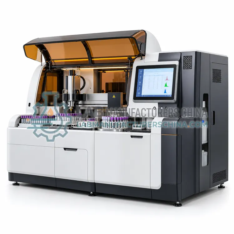
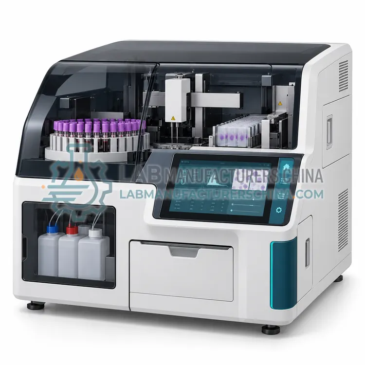
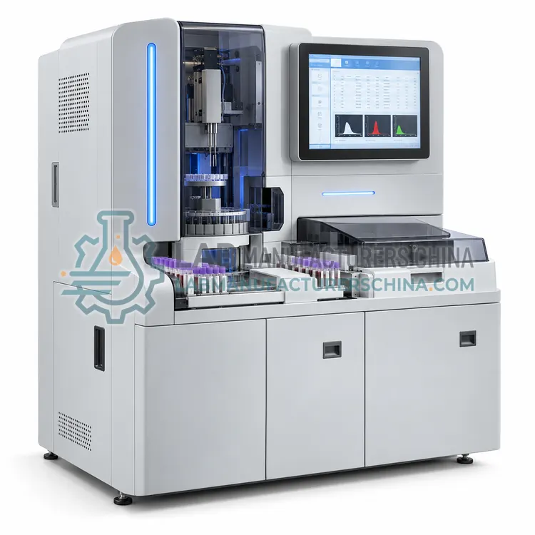
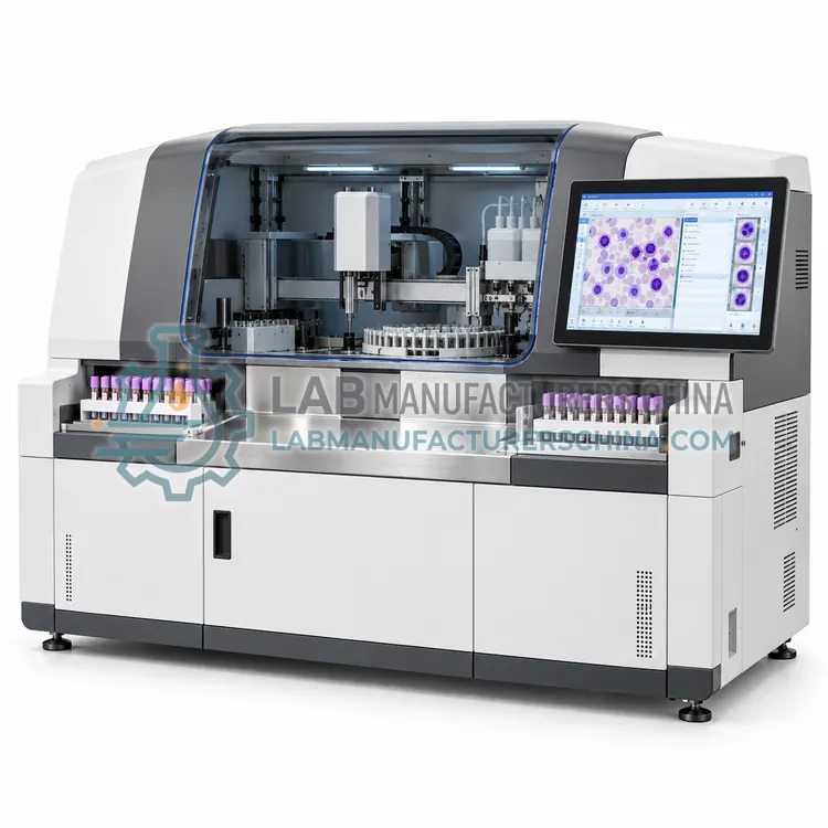
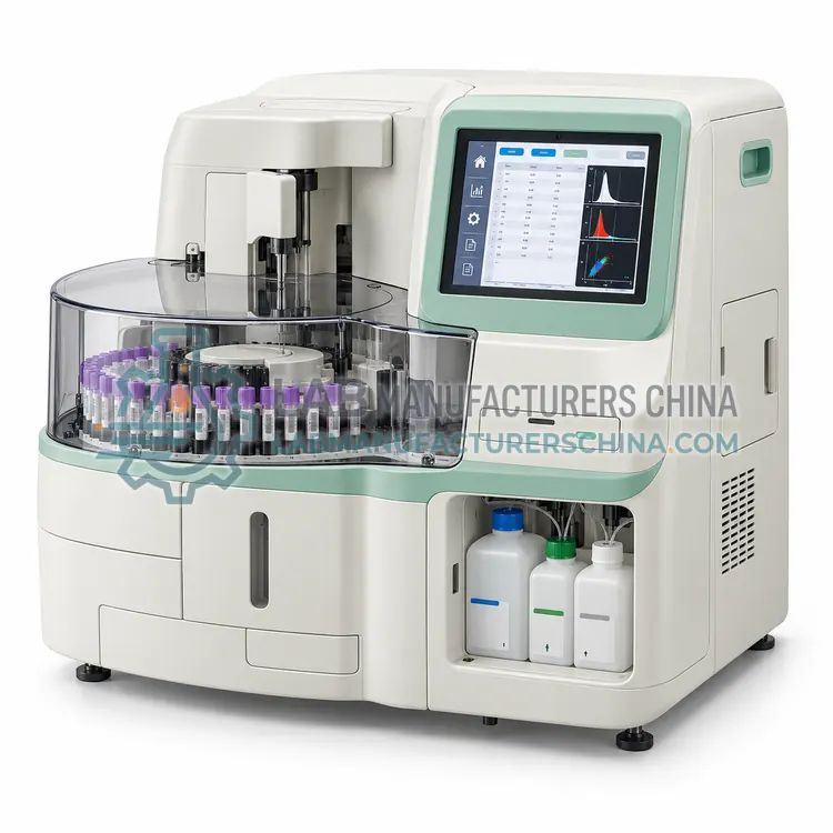
An automated hematology system is an assembly of modules rather than a single instrument. The analytical module performs impedance counting and laser flow cytometry to produce the complete blood count and five-part differential. Pre-analytical elements include a bulk loader, a centrifuge or mixing station, and a barcode-driven sorter that directs tubes according to their test orders. Post-analytical elements include a slide maker and stainer that prepares films for samples meeting defined review criteria, a digital morphology scanner that takes images of the films, and middleware that applies review rules, routes work between instruments and manages quality control across the system. Communication uses barcode identification and HL7 or ASTM interfaces to the laboratory information system, so that a tube is measured, reviewed and reported with minimal manual handling, while the middleware records which rules were applied to each result.
Which Automated Hematology Systems variants are available?
- Track-Connected Automated Hematology System — A track-connected hematology system links analyzers, loaders and review stations to a laboratory conveyor, so tubes arriving from the core laboratory are routed, measured and reported automatically.
- Automated Hematology Analyzer with Slide Maker and Stainer — This system pairs a five-part analyzer with an onboard slide maker and stainer that prepares and stains films automatically when review criteria are met.
- Modular Automated Hematology Workcell — A modular hematology workcell groups two or more analyzers under shared middleware so that workload can be split, backed up or scaled in steps.
- Automated Digital Cell Morphology System — A digital cell morphology system scans stained blood films, locates and images cells with automated recognition, and presents them to a technologist for classification.
- Automated Hematology Analyzer with Integrated Body Fluid Module — This analyzer adds a body fluid mode that counts cells in cerebrospinal, pleural, peritoneal and synovial fluid using the same flow cytometry channel as the blood count.
How do I choose the right variant?
Choose an automated hematology system when daily counts exceed several hundred, when film review consumes significant technologist time, or when the laboratory wants to consolidate hematology with other disciplines on a track. Compare systems by which modules are integrated, whether film preparation is triggered automatically by analyzer flags, whether digital images are stored for remote review, and how middleware handles reflex rules. Check throughput of the loader and the analyzer together, because the slower module sets the pace. Confirm reagent and consumable logistics for each module, the floor space and service clearances required, the validation workload at installation, and whether the supplier can support middleware changes locally. Plan the workflow and review rules before purchase, not after.
Where are Automated Hematology Systems used?
Automated Hematology Systems are specified in high-volume hospital hematology laboratories, consolidated core laboratory workflows, reference laboratories with remote morphology review and multi-site laboratory networks with middleware rules.
Which standards and certifications apply?
Work with Automated Hematology Systems is commonly governed by ISO 15189, CLSI H26-A2, CLSI AUTO10, CLSI EP05, CLSI H20-A2, IEC 61010-1 and IEC 61010-2-101. Instrument certifications such as CE marking are confirmed per configuration and never assumed.
What are the key considerations before purchase?
Installation requires a stable environment with controlled temperature, low dust, adequate floor loading for track sections and uninterruptible power for the controller and middleware server. Verify each module separately before validating the integrated workflow, including barcode read rates, routing accuracy, slide quality and image capture. Document review rules and test them against a representative sample set before go-live, because rule errors affect patient results. Plan daily cleaning of probes and tracks, weekly flow cell and optics checks, slide stainer reagent replacement and periodic replacement of pneumatic parts. Keep a downtime procedure that allows analysis to continue on a stand-alone analyzer if the track or middleware fails, and treat all samples, films and waste as biohazardous.