Automated Digital Cell Morphology System
自动数字细胞形态分析系统
- SKU AHS-0004
- Automated Hematology Systems
- CE (to be confirmed)
- ISO 13485 manufacturing (to be confirmed)
- NMPA class II registration (to be confirmed)
Price on request — FOB / CIF quoted per order
Prices depend on configuration, quantity and destination port.
Key specifications
| Scanning capacity | 20–40 slides/h depending on cell count |
|---|---|
| Cell images per slide | Up to 200 white cell images |
| Image resolution | 0.05–0.1 µm per pixel |
| Pre-classification categories | Five-part differential plus abnormal cell classes |
| Stain compatibility | Romanowsky-type stains |
| Data storage | Images and results archived on server, ≥1 TB |
Request a quote Download brochure · to be provided
By Lab Manufacturers China Technical TeamLast reviewed Standards: ISO 15189, CLSI H26-A2, CLSI AUTO10
What is the Automated Digital Cell Morphology System?
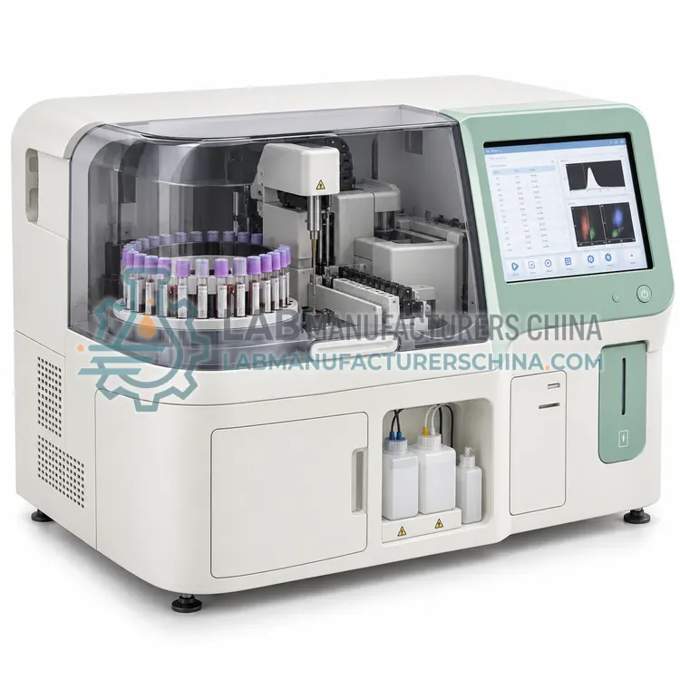
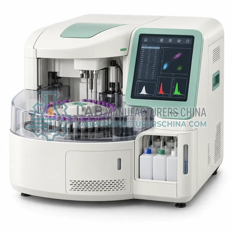
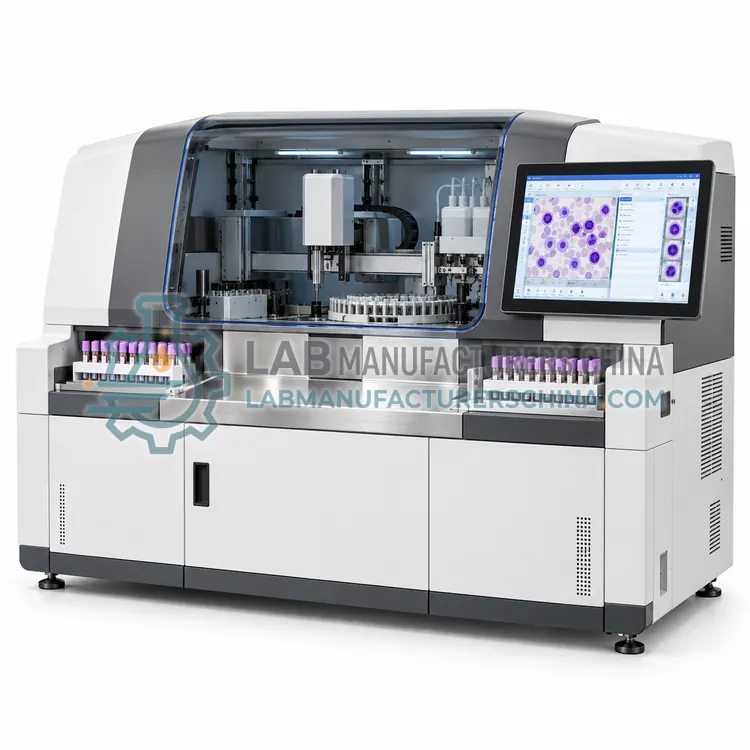
A digital cell morphology system scans stained blood films, locates and images cells with automated recognition, and presents them to a technologist for classification. It suits laboratories that need consistent film examination with image archiving and remote review.
What is the Automated Digital Cell Morphology System?
A digital cell morphology system scans stained blood films, locates and images cells with automated recognition, and presents them to a technologist for classification. It suits laboratories that need consistent film examination with image archiving and remote review.
Overview
The system combines a slide scanner with image analysis software. A stained film is loaded, the analyzer locates the monolayer, captures high-resolution images of white cells, red cells and platelets, and pre-classifies them using pattern recognition trained on reference libraries. The technologist reviews the pre-classified images on screen, corrects any misclassification, and issues the differential, which is then sent to the laboratory information system. Because images and decisions are stored, the system supports retrospective review, teaching and quality audits. Some platforms also perform automatic red cell morphology scoring and platelet estimates. Turnaround depends on the number of cells examined, typically five to ten minutes per film, and image quality depends on correct film preparation and staining.
How does it work?
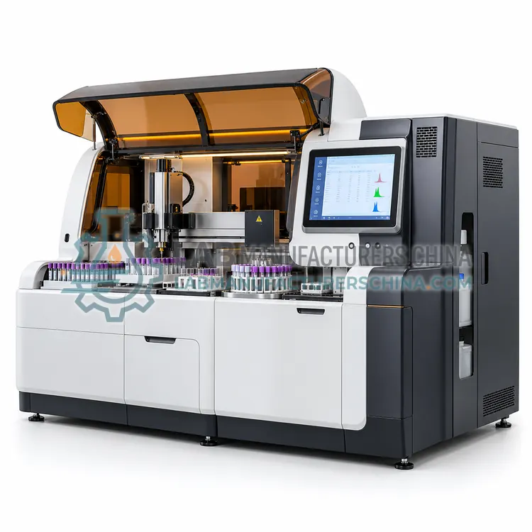
An automated hematology system is an assembly of modules rather than a single instrument. The analytical module performs impedance counting and laser flow cytometry to produce the complete blood count and five-part differential. Pre-analytical elements include a bulk loader, a centrifuge or mixing station, and a barcode-driven sorter that directs tubes according to their test orders. Post-analytical elements include a slide maker and stainer that prepares films for samples meeting defined review criteria, a digital morphology scanner that takes images of the films, and middleware that applies review rules, routes work between instruments and manages quality control across the system. Communication uses barcode identification and HL7 or ASTM interfaces to the laboratory information system, so that a tube is measured, reviewed and reported with minimal manual handling, while the middleware records which rules were applied to each result.
What are the key features?
- Automated slide scanning with high-resolution cell imaging
- Pre-classification of white cells using trained pattern recognition
- On-screen review and correction workstations for technologists
- Archived images linked to the patient result and audit trail
- Automatic red cell morphology scoring and platelet estimation
- Remote review through a network workstation option
What are the technical specifications?
A typical automated digital cell morphology system configuration offers scanning capacity of 20–40 slides/h depending on cell count, cell images per slide of Up to 200 white cell images, image resolution of 0.05–0.1 µm per pixel, pre-classification categories of Five-part differential plus abnormal cell classes, stain compatibility of Romanowsky-type stains and data storage of Images and results archived on server, ≥1 TB. Values are indicative of the product class; the exact figures for each configuration are confirmed on the quotation and the calibration documents supplied with the instrument.
Which applications is it used for?
The Automated Digital Cell Morphology System is used in hematology film review in hospital laboratories, remote morphology review for satellite sites, teaching hospital case archives and quality audit of differential accuracy. Across the wider Automated Hematology Systems class, laboratories also deploy it for high-volume hospital hematology laboratories, consolidated core laboratory workflows and reference laboratories with remote morphology review.
Which standards and certifications apply?
Relevant standards and methods include ISO 15189, CLSI H26-A2, CLSI AUTO10, CLSI EP05, CLSI H20-A2, IEC 61010-1 and IEC 61010-2-101. Certification candidates for this model are CE, ISO 13485 manufacturing and NMPA class II registration; each is to be confirmed for the specific configuration before shipment, and certificates are supplied only where they have been issued.
Installation requires a stable environment with controlled temperature, low dust, adequate floor loading for track sections and uninterruptible power for the controller and middleware server. Verify each module separately before validating the integrated workflow, including barcode read rates, routing accuracy, slide quality and image capture. Document review rules and test them against a representative sample set before go-live, because rule errors affect patient results. Plan daily cleaning of probes and tracks, weekly flow cell and optics checks, slide stainer reagent replacement and periodic replacement of pneumatic parts. Keep a downtime procedure that allows analysis to continue on a stand-alone analyzer if the track or middleware fails, and treat all samples, films and waste as biohazardous.
How do I select the right Automated Digital Cell Morphology System?
Select a digital morphology system when film review volume is high enough to justify it, typically more than about thirty films per day, or when remote review, archiving or teaching is required. In the RFQ ask about the accuracy of pre-classification, the technologist time needed per film, storage requirements, and integration with your hematology analyzers and slide maker. Request a side-by-side comparison with manual microscopy during evaluation.
How does it compare with other Automated Hematology Systems variants?
Choose an automated hematology system when daily counts exceed several hundred, when film review consumes significant technologist time, or when the laboratory wants to consolidate hematology with other disciplines on a track. Compare systems by which modules are integrated, whether film preparation is triggered automatically by analyzer flags, whether digital images are stored for remote review, and how middleware handles reflex rules. Check throughput of the loader and the analyzer together, because the slower module sets the pace. Confirm reagent and consumable logistics for each module, the floor space and service clearances required, the validation workload at installation, and whether the supplier can support middleware changes locally. Plan the workflow and review rules before purchase, not after.
Full specifications
| Scanning capacity | 20–40 slides/h depending on cell count |
|---|---|
| Cell images per slide | Up to 200 white cell images |
| Image resolution | 0.05–0.1 µm per pixel |
| Pre-classification categories | Five-part differential plus abnormal cell classes |
| Stain compatibility | Romanowsky-type stains |
| Data storage | Images and results archived on server, ≥1 TB |
| Review workstations | Up to 5 networked workstations |
| Interfaces | HL7 or ASTM to LIS; DICOM optional |
| Power supply | AC 100–240 V, 50/60 Hz |
Indicative values for this product class. Final specifications are confirmed on the quotation and in the calibration documents supplied with the instrument.
Applications
- Hematology film review in hospital laboratories
- Remote morphology review for satellite sites
- Teaching hospital case archives
- Quality audit of differential accuracy
Key features
- Automated slide scanning with high-resolution cell imaging
- Pre-classification of white cells using trained pattern recognition
- On-screen review and correction workstations for technologists
- Archived images linked to the patient result and audit trail
- Automatic red cell morphology scoring and platelet estimation
- Remote review through a network workstation option
Frequently asked questions about Automated Digital Cell Morphology System
It is normally positioned as a review aid: the technologist remains responsible for the final differential, and the system pre-classifies and presents images. Regulatory classification depends on the market and on the claims made, so confirm how the device is registered in your jurisdiction before purchase.
The reference method assesses 100 white cells for a standard differential, with more cells examined when the count is low or abnormal populations are present. Because the system stores many more images than are reviewed, laboratories can review additional cells without preparing another slide.
Image archives grow quickly, typically several gigabytes per thousand films, so a dedicated server with at least one terabyte of usable capacity and a defined retention policy is advisable. Many laboratories keep full images for a limited period and retain only summary images after that.
A typical configuration includes a loader or track connection, one or two hematology analyzers, a slide maker and stainer, optionally a digital morphology scanner, and middleware that applies review rules and routes samples. Centrifuges and sorters may be added when the system is part of a wider core laboratory track.
No, but it changes it. Automated systems prepare and stain films on demand, and digital scanning can present images to a technologist at a remote workstation. The medical decision to review morphology and the interpretation of the film remain human tasks, guided by documented rules agreed with the clinical service.
Each module is verified individually for precision, carryover and accuracy, and the integrated workflow is then validated for barcode reading, routing, film quality and data flow to the LIS. The laboratory also tests its review rules on a representative set of normal and abnormal samples, including known abnormal cases.
Related models
Automated Hematology Analyzer with Integrated Body Fluid Module
带集成体液模块的自动化血液分析仪
- CE (to be confirmed)
- +2