Automated Cytology Slide Preparation System
自动细胞学制片系统
- SKU SPS-0001
- Slide Preparation Systems
- CE (to be confirmed)
- IEC 61010-1 compliance (to be confirmed)
- ISO 13485 manufacturing (to be confirmed)
Price on request — FOB / CIF quoted per order
Prices depend on configuration, quantity and destination port.
Key specifications
| Samples per run | 24–48 vials |
|---|---|
| Spot diameter | about 13–20 mm |
| Cycle time | about 30–60 min per batch |
| Traceability | Barcode on vial and slide |
| Integration | Drying, staining, coverslipping options |
| Reagent supply | On-board with level sensing |
Request a quote Download brochure · to be provided
By Lab Manufacturers China Technical TeamLast reviewed Standards: IEC 61010-1, IEC 61010-2-101, IEC 61326-1
What is the Automated Cytology Slide Preparation System?
An automated cytology slide preparation system performs sample dilution, deposition and slide processing without manual intervention. It is chosen by high-volume screening and diagnostic laboratories that need standardised, traceable and highly reproducible preparations.
What is the Automated Cytology Slide Preparation System?
An automated cytology slide preparation system performs sample dilution, deposition and slide processing without manual intervention. It is chosen by high-volume screening and diagnostic laboratories that need standardised, traceable and highly reproducible preparations.
Overview
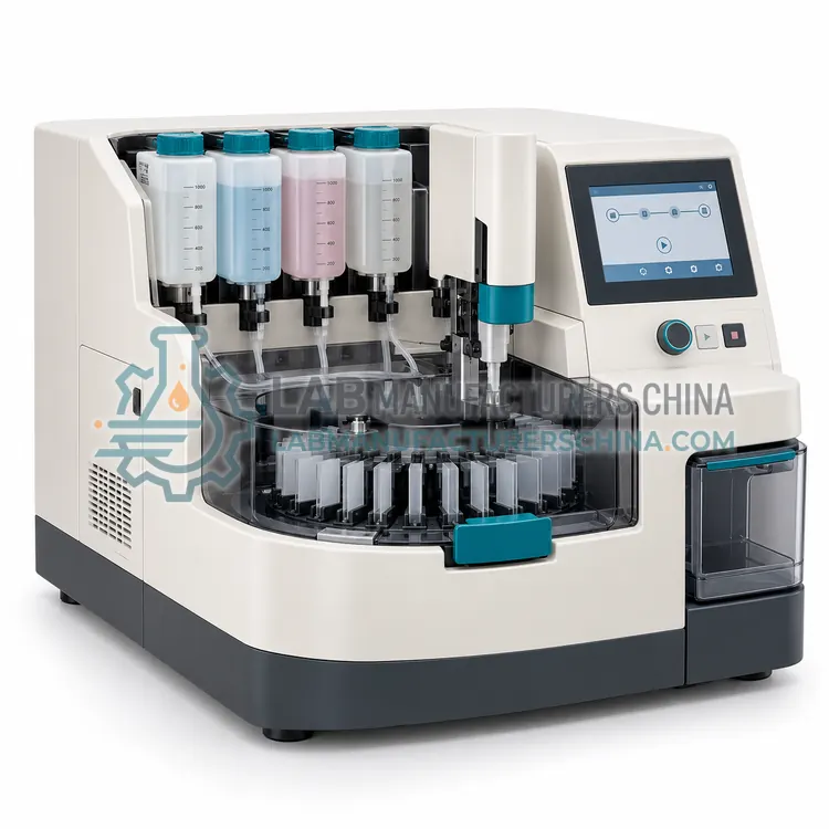
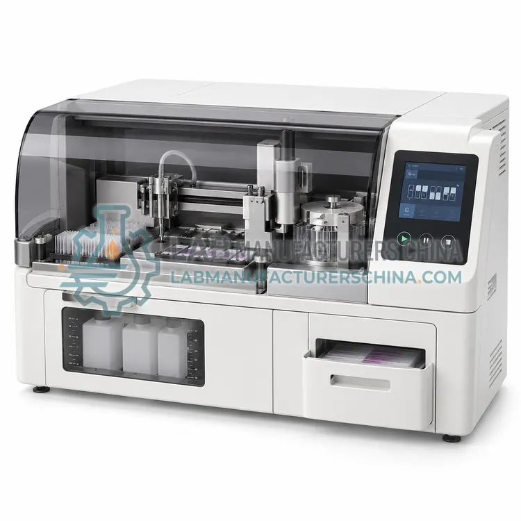
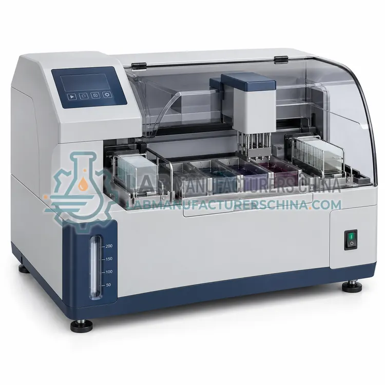
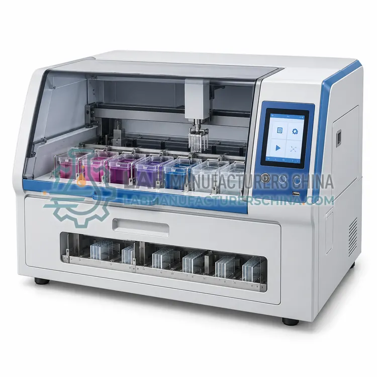
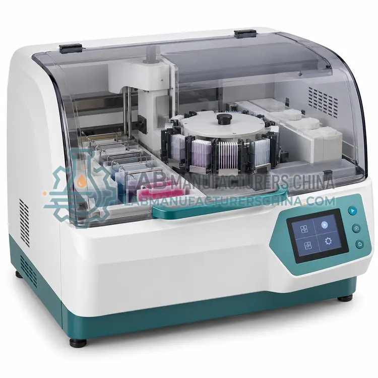
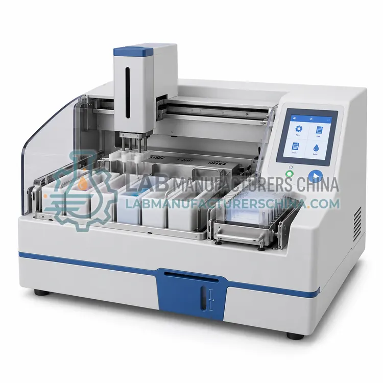
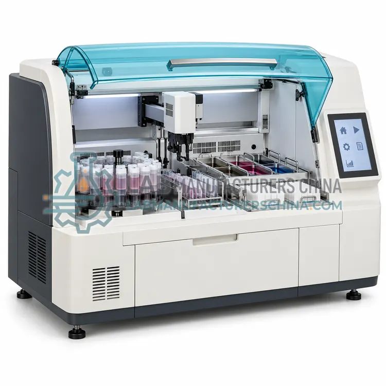
The automated cytology slide preparation system links sample handling, deposition and downstream slide processing under one control system. Barcoded vials are loaded, the instrument dilutes and mixes the sample, deposits a measured aliquot onto a slide, and moves it through drying and often staining and coverslipping. Operating parameters are stored as protocols, so different sample types receive appropriate treatment without operator decisions. Record keeping is a major benefit: the system can log which sample produced which slide and when. Throughput is typically tens of samples per run, and some platforms run continuously. The main requirements are consistent sample collection, adequate cell numbers, correct reagent supply and scheduled cleaning, because a blockage or reagent shortage can affect an entire batch.
How does it work?
A slide preparation system combines sample handling with deposition and, often, downstream slide processing. Depending on the design, it may dilute and mix the sample, deposit a measured volume onto a slide, spread or settle cells into a defined area, dry the slide, transfer it to a stainer and finally apply a coverslip. Many systems use barcode tracking so each sample, slide and reagent event is linked in a record. Motors, syringes or peristaltic pumps move fluid, and heated or ventilated zones control drying. Consistency comes from fixed volumes, timings and motions, which reduce operator-dependent variation. The quality of the output depends on correct sample collection, adequate cell numbers and complete reagent removal, because residual fluid or fixative causes uneven staining and poor adhesion.
What are the key features?
- Barcoded sample loading with traceability
- Automated dilution, mixing and measured deposition
- Integrated drying, staining and coverslipping options
- Stored protocols for different sample types
- Batch or continuous operation
- Run logs linking samples to slides
What are the technical specifications?
A typical automated cytology slide preparation system configuration offers samples per run of 24–48 vials, spot diameter of about 13–20 mm, cycle time of about 30–60 min per batch, traceability of Barcode on vial and slide, integration of Drying, staining, coverslipping options and reagent supply of On-board with level sensing. Values are indicative of the product class; the exact figures for each configuration are confirmed on the quotation and the calibration documents supplied with the instrument.
Which applications is it used for?
The Automated Cytology Slide Preparation System is used in high-volume cervical screening, diagnostic cytology services, body fluid cytology and laboratories with traceability requirements. Across the wider Slide Preparation Systems class, laboratories also deploy it for cytology slide preparation, cervical screening programmes and body fluid cytology.
Which standards and certifications apply?
Relevant standards and methods include IEC 61010-1, IEC 61010-2-101, IEC 61326-1, ISO 15189 and CLSI GP41. Certification candidates for this model are CE, IEC 61010-1 compliance and ISO 13485 manufacturing; each is to be confirmed for the specific configuration before shipment, and certificates are supplied only where they have been issued.
Install the system on a stable bench with power and, if required, a drain, and keep the working area organised so tubes and slides cannot be mismatched. Follow the specified reagent volumes and timings, since too little fixative causes poor adhesion and too much leaves residue. Clean fluid lines and nozzles on schedule to prevent blockage and carryover, and check that barcodes are readable so records remain complete. Verify performance with control samples and record reagent lot numbers and run conditions. Train staff in safe handling of preservative fluids and in recognising preparation faults, and keep maintenance and quality records for accreditation.
How do I select the right Automated Cytology Slide Preparation System?
Specify a fully automated system for high-volume work where standardisation and traceability are essential. Confirm samples per run, cycle time, integration options, barcode architecture and consumable cost per slide. Plan reagent management and service contracts, since an interruption affects an entire batch.
How does it compare with other Slide Preparation Systems variants?
Choose a preparation system by the level of automation you need and the output you must produce. A semi-automated system suits laboratories that want control over sample selection while automating deposition. A fully automated system gives standardised monolayers at throughput, often with integrated staining and coverslipping. Liquid-based systems define the reagent and consumable architecture, while cell block systems target ancillary testing. Compare samples per run, cycle time, spot size, barcode and record capability, consumable cost and compatibility with existing stainers. Check whether the system is open to third-party reagents, and confirm validation, training and service support before committing to a platform.
Full specifications
| Samples per run | 24–48 vials |
|---|---|
| Spot diameter | about 13–20 mm |
| Cycle time | about 30–60 min per batch |
| Traceability | Barcode on vial and slide |
| Integration | Drying, staining, coverslipping options |
| Reagent supply | On-board with level sensing |
| Slide format | 25 × 75 mm standard |
| Power supply | 220 V, 50 Hz, about 1 kW |
Indicative values for this product class. Final specifications are confirmed on the quotation and in the calibration documents supplied with the instrument.
Applications
- High-volume cervical screening
- Diagnostic cytology services
- Body fluid cytology
- Laboratories with traceability requirements
Key features
- Barcoded sample loading with traceability
- Automated dilution, mixing and measured deposition
- Integrated drying, staining and coverslipping options
- Stored protocols for different sample types
- Batch or continuous operation
- Run logs linking samples to slides
Frequently asked questions about Automated Cytology Slide Preparation System
Depending on batch size and cycle time, a fully automated system can prepare several hundred samples per shift, often running continuously. Actual output also depends on staining and coverslipping throughput downstream.
No. Staff still evaluate sample adequacy, load and unload vials correctly, monitor reagent levels and interpret control results. The system reduces repetitive handling and timing variation, not the need for trained judgement.
Most systems flag the vial and pause or divert it, so the sample is not processed without identity. Investigate label quality and printing, and consider re-labelling with a verified barcode before it is loaded again.
It automates fluid handling and cell deposition, and often drying, staining and coverslipping. Sample dilution, mixing, measured deposition and barcode tracking are typical functions, so the output is a standardised, traceable slide ready for microscopy.
Only if daily volume is high or standardisation is critical. Semi-automated systems that automate deposition while leaving sample selection to the operator often suit moderate volumes and cost considerably less, with simpler maintenance.
Through barcoded sample vials and slides linked in the instrument software, so every preparation step is recorded against the correct specimen. Manual systems rely on labels and written logs, which are more error-prone at high volume.