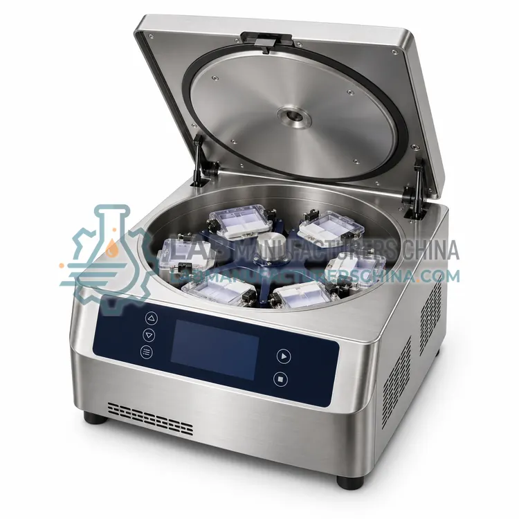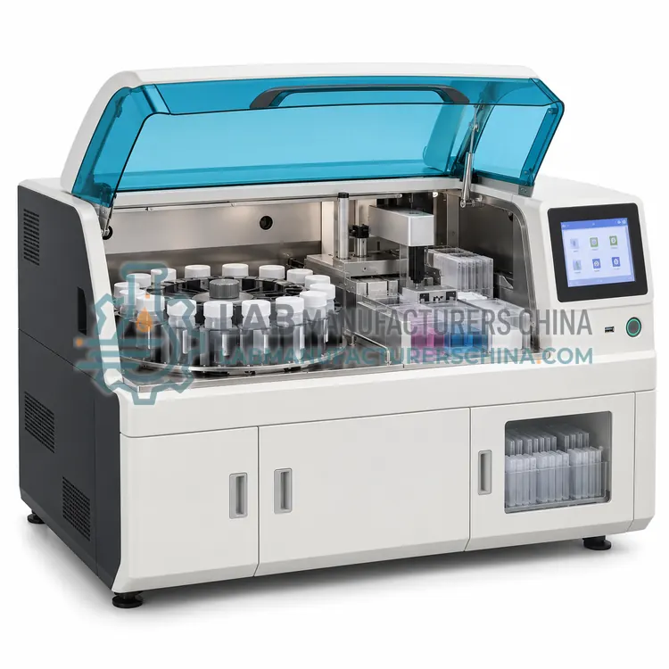
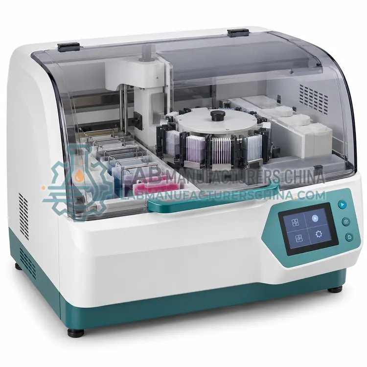
Thin-Layer Cytology Processor
薄层细胞学处理机
- SKU CYP-0002
- Cytology Processors
- CE (to be confirmed)
- IEC 61010-1 compliance (to be confirmed)
- ISO 13485 manufacturing (to be confirmed)
Price on request — FOB / CIF quoted per order
Prices depend on configuration, quantity and destination port.
Key specifications
| Samples per run | 1–24 vials |
|---|---|
| Spot diameter | about 10–15 mm |
| Cell density control | Measured volume or counted cells |
| Preparation thickness | Single cell layer |
| Cycle time | about 20–45 min per batch |
| Slide format | 25 × 75 mm standard |
Request a quote Download brochure · to be provided
By Lab Manufacturers China Technical TeamLast reviewed Standards: IEC 61010-1, IEC 61010-2-101, IEC 61326-1
What is the Thin-Layer Cytology Processor?
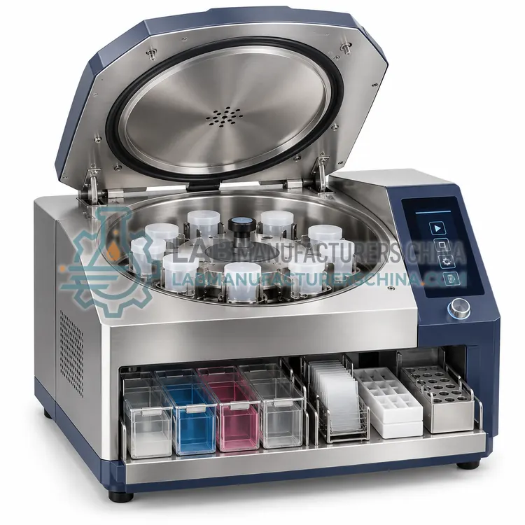
A thin-layer cytology processor deposits a controlled-density cell layer on a slide using a spreading or settling technique. It is chosen when a thin, consistent preparation is needed for screening and image analysis.
What is the Thin-Layer Cytology Processor?
A thin-layer cytology processor deposits a controlled-density cell layer on a slide using a spreading or settling technique. It is chosen when a thin, consistent preparation is needed for screening and image analysis.
Overview
Thin-layer preparation aims for a uniform, controlled thickness of cells rather than a random smear. The processor may spread a measured volume of cell suspension over a defined area, allow cells to settle by gravity or gentle centrifugation, or use a filter transfer to deposit a thin film. Density is regulated by the cell concentration in the suspension and the volume applied, so the instrument often estimates or standardises cell numbers. The result is a preparation where cells lie in a single plane, which improves microscopic examination and is well suited to automated image analysis and digital cytology. Thin-layer methods are used in gynaecological screening, urine cytology and some body fluids, and can be combined with cell block preparation for ancillary testing. Consistency across operators is the main benefit.
How does it work?
A cytology processor starts with a sample collected into a preservative solution, which fixes cells and lyses red blood cells. The instrument then separates diagnostically useful cells from mucus, blood and inflammatory debris using one of several approaches. Density gradient or centrifugation methods layer the sample over a reagent so cells of interest band at a known density. Filtration and membrane methods trap cells on a surface with a defined pore size, then transfer them to a slide. Some platforms use an adhesive or charged coating to capture cells while washing away the rest. The result is a thin, evenly distributed preparation with cells concentrated in a defined area, which improves screening speed and reduces obscuring material. Cell recovery, spot area and carryover control determine performance.
What are the key features?
- Measured cell suspension spread over a defined area
- Controlled cell density and preparation thickness
- Single-plane cell distribution
- Suited to automated image analysis
- Compatible with cell block preparation
- Consistent results between operators
What are the technical specifications?
A typical thin-layer cytology processor configuration offers samples per run of 1–24 vials, spot diameter of about 10–15 mm, cell density control of Measured volume or counted cells, preparation thickness of Single cell layer, cycle time of about 20–45 min per batch and slide format of 25 × 75 mm standard. Values are indicative of the product class; the exact figures for each configuration are confirmed on the quotation and the calibration documents supplied with the instrument.
Which applications is it used for?
The Thin-Layer Cytology Processor is used in gynaecological cytology screening, urine cytology, body fluid cytology and digital and automated image analysis. Across the wider Cytology Processors class, laboratories also deploy it for cervical cancer screening, urine cytology and body fluid cytology.
Which standards and certifications apply?
Relevant standards and methods include IEC 61010-1, IEC 61010-2-101, IEC 61326-1, ISO 15189 and CLSI GP41. Certification candidates for this model are CE, IEC 61010-1 compliance and ISO 13485 manufacturing; each is to be confirmed for the specific configuration before shipment, and certificates are supplied only where they have been issued.
Place the processor on a stable bench near a drain and power outlet, and keep the working area free of clutter so sample tubes can be tracked. Follow the specified reagent volumes and incubation or centrifugation conditions, because under-processing leaves debris while over-processing loses cells. Change reagents and clean fluid paths on schedule to prevent contamination and biofilm. Verify performance with control samples and record reagent lot numbers, run dates and results. Train staff in safe handling of preservative fluids, which may contain methanol or other hazardous substances, and dispose of waste according to local regulations.
How do I select the right Thin-Layer Cytology Processor?
Select a thin-layer processor when uniform, single-plane preparations are needed for screening or image analysis, or when cell density must be standardised across samples. Confirm spot size, density control method, cycle time and compatibility with your staining and imaging workflow.
How does it compare with other Cytology Processors variants?
Choose a processor by sample type, throughput and whether the laboratory prefers an open or closed reagent system. For high-volume cervical screening, a liquid-based processor with automated staining and coverslipping gives the most consistent results. For body fluids and fine-needle aspirates, a centrifugation or filter-based method often yields better cell recovery from scant samples. Cell block processors support immunohistochemistry and molecular testing on residual cells. Compare samples per run, reagent consumption, carryover prevention, consumable cost and whether the platform accepts third-party reagents. Confirm validation, training and service support, and check that the output format fits existing stainers and coverslippers.
Full specifications
| Samples per run | 1–24 vials |
|---|---|
| Spot diameter | about 10–15 mm |
| Cell density control | Measured volume or counted cells |
| Preparation thickness | Single cell layer |
| Cycle time | about 20–45 min per batch |
| Slide format | 25 × 75 mm standard |
| Staining | Compatible with Papanicolaou staining |
| Power supply | 220 V, 50 Hz, about 0.4 kW |
Indicative values for this product class. Final specifications are confirmed on the quotation and in the calibration documents supplied with the instrument.
Applications
- Gynaecological cytology screening
- Urine cytology
- Body fluid cytology
- Digital and automated image analysis
Key features
- Measured cell suspension spread over a defined area
- Controlled cell density and preparation thickness
- Single-plane cell distribution
- Suited to automated image analysis
- Compatible with cell block preparation
- Consistent results between operators
Frequently asked questions about Thin-Layer Cytology Processor
Cells lie in a single plane at controlled density, so they are easier to examine and less likely to overlap or be obscured. Screening is faster, and the preparation is well suited to automated image analysis and archiving.
By measuring the volume of cell suspension applied, by counting or estimating cell concentration, or by adjusting settling conditions. Consistent density between samples makes screening effort more predictable and supports automated analysis.
Yes, prepared slides can be used for immunocytochemistry or in-situ hybridisation, and residual cells can be processed into a cell block for further immunohistochemical or molecular testing. This preserves the original screening preparation.
It prepares a cell sample from liquid suspension into a slide-ready monolayer. It removes mucus, blood and debris, concentrates diagnostically useful cells and deposits them in a defined area so the microscopist can screen the slide quickly and reliably.
The sample is rinsed into a preservative fluid rather than smeared directly onto a slide. The processor then removes debris and deposits a representative cell population as a thin monolayer, which usually improves cell distribution and reduces obscuring blood and mucus.
Filter methods trap cells on a membrane with a defined pore size and transfer them to a slide, while centrifugation methods concentrate cells by density. Filtration generally gives cleaner preparations, and centrifugation often recovers more cells from very scant samples.
Related models