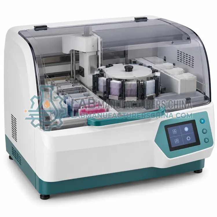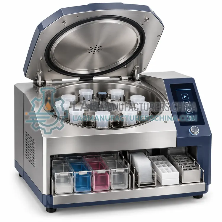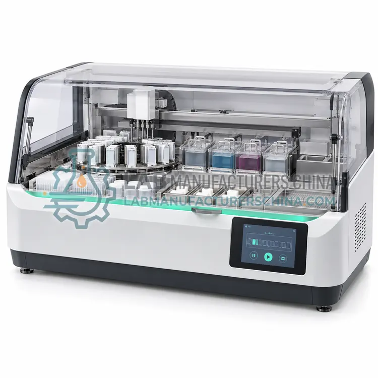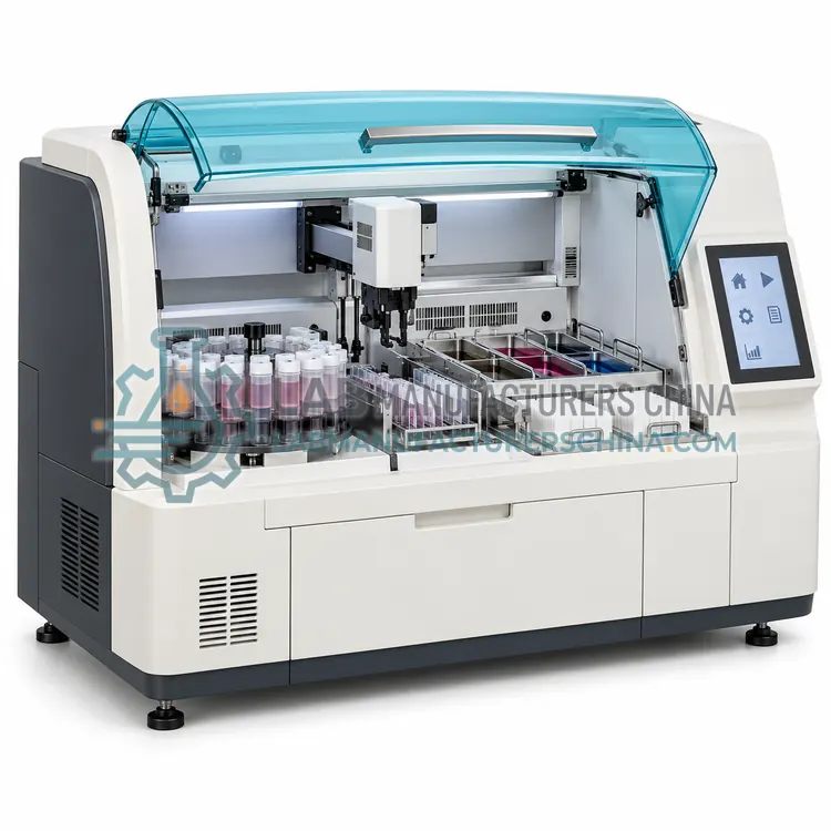
Centrifugation-Based Cytology Processor
离心式细胞学处理机
- SKU CYP-0005
- Cytology Processors
- CE (to be confirmed)
- IEC 61010-1 compliance (to be confirmed)
- ISO 13485 manufacturing (to be confirmed)
Price on request — FOB / CIF quoted per order
Prices depend on configuration, quantity and destination port.
Key specifications
| Separation principle | Density gradient or differential spin |
|---|---|
| Spin speed | 200–3000 rpm |
| Cycle time | about 5–20 min |
| Minimum sample volume | about 0.2–1 mL |
| Spot diameter | about 6–13 mm |
| Cell recovery | High for low-cellularity fluids |
Request a quote Download brochure · to be provided
By Lab Manufacturers China Technical TeamLast reviewed Standards: IEC 61010-1, IEC 61010-2-101, IEC 61326-1
What is the Centrifugation-Based Cytology Processor?
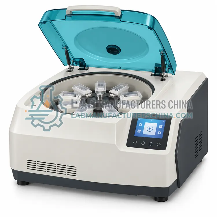
A centrifugation-based cytology processor concentrates cells by density, often through a gradient, to prepare slides from fluids that contain few cells. It is chosen for body fluids, cerebrospinal fluid and aspirate rinses.
What is the Centrifugation-Based Cytology Processor?
A centrifugation-based cytology processor concentrates cells by density, often through a gradient, to prepare slides from fluids that contain few cells. It is chosen for body fluids, cerebrospinal fluid and aspirate rinses.
Overview
Centrifugation-based preparation relies on differences in density to separate cells from fluid and debris. The sample may be layered over a density gradient medium so that cells of interest collect at an interface while lighter debris stays above and denser material sinks. Alternatively, a simple differential spin pellets cells that are then resuspended at a controlled concentration before deposition on a slide. Compared with filtration, centrifugation usually recovers more cells from sparse samples and handles samples with very few cells better, which suits cerebrospinal fluid, serous effusions and aspirate rinses. Cell clumping and protein background can occur if conditions are not optimised, so spin speed, time and medium density matter. Some processors combine centrifugation with a cytospin-style deposition step for a defined spot on the slide.
How does it work?
A cytology processor starts with a sample collected into a preservative solution, which fixes cells and lyses red blood cells. The instrument then separates diagnostically useful cells from mucus, blood and inflammatory debris using one of several approaches. Density gradient or centrifugation methods layer the sample over a reagent so cells of interest band at a known density. Filtration and membrane methods trap cells on a surface with a defined pore size, then transfer them to a slide. Some platforms use an adhesive or charged coating to capture cells while washing away the rest. The result is a thin, evenly distributed preparation with cells concentrated in a defined area, which improves screening speed and reduces obscuring material. Cell recovery, spot area and carryover control determine performance.
What are the key features?
- Density gradient separation of cells from debris
- High cell recovery from sparse samples
- Resuspension to a controlled cell concentration
- Suitable for cerebrospinal fluid and effusions
- Deposition onto a defined slide spot
- Adjustable spin speed and time
What are the technical specifications?
A typical centrifugation-based cytology processor configuration offers separation principle of Density gradient or differential spin, spin speed of 200–3000 rpm, cycle time of about 5–20 min, minimum sample volume of about 0.2–1 mL, spot diameter of about 6–13 mm and cell recovery of High for low-cellularity fluids. Values are indicative of the product class; the exact figures for each configuration are confirmed on the quotation and the calibration documents supplied with the instrument.
Which applications is it used for?
The Centrifugation-Based Cytology Processor is used in cerebrospinal fluid cytology, serous effusion preparation, fine-needle aspirate rinses and low-cellularity body fluids. Across the wider Cytology Processors class, laboratories also deploy it for cervical cancer screening, urine cytology and body fluid cytology.
Which standards and certifications apply?
Relevant standards and methods include IEC 61010-1, IEC 61010-2-101, IEC 61326-1, ISO 15189 and CLSI GP41. Certification candidates for this model are CE, IEC 61010-1 compliance and ISO 13485 manufacturing; each is to be confirmed for the specific configuration before shipment, and certificates are supplied only where they have been issued.
Place the processor on a stable bench near a drain and power outlet, and keep the working area free of clutter so sample tubes can be tracked. Follow the specified reagent volumes and incubation or centrifugation conditions, because under-processing leaves debris while over-processing loses cells. Change reagents and clean fluid paths on schedule to prevent contamination and biofilm. Verify performance with control samples and record reagent lot numbers, run dates and results. Train staff in safe handling of preservative fluids, which may contain methanol or other hazardous substances, and dispose of waste according to local regulations.
How do I select the right Centrifugation-Based Cytology Processor?
Choose centrifugation-based preparation when samples are sparse and cell recovery matters more than background cleanliness, as with cerebrospinal fluid and effusions. Confirm minimum sample volume, spin speed range, gradient or differential options and deposition format. Check recovery with your own low-cellularity samples.
How does it compare with other Cytology Processors variants?
Choose a processor by sample type, throughput and whether the laboratory prefers an open or closed reagent system. For high-volume cervical screening, a liquid-based processor with automated staining and coverslipping gives the most consistent results. For body fluids and fine-needle aspirates, a centrifugation or filter-based method often yields better cell recovery from scant samples. Cell block processors support immunohistochemistry and molecular testing on residual cells. Compare samples per run, reagent consumption, carryover prevention, consumable cost and whether the platform accepts third-party reagents. Confirm validation, training and service support, and check that the output format fits existing stainers and coverslippers.
Full specifications
| Separation principle | Density gradient or differential spin |
|---|---|
| Spin speed | 200–3000 rpm |
| Cycle time | about 5–20 min |
| Minimum sample volume | about 0.2–1 mL |
| Spot diameter | about 6–13 mm |
| Cell recovery | High for low-cellularity fluids |
| Slide format | 25 × 75 mm standard |
| Power supply | 220 V, 50 Hz, about 0.3 kW |
Indicative values for this product class. Final specifications are confirmed on the quotation and in the calibration documents supplied with the instrument.
Applications
- Cerebrospinal fluid cytology
- Serous effusion preparation
- Fine-needle aspirate rinses
- Low-cellularity body fluids
Key features
- Density gradient separation of cells from debris
- High cell recovery from sparse samples
- Resuspension to a controlled cell concentration
- Suitable for cerebrospinal fluid and effusions
- Deposition onto a defined slide spot
- Adjustable spin speed and time
Frequently asked questions about Centrifugation-Based Cytology Processor
For samples with very few cells, because filtration can lose cells on the membrane and in the filter housing. Centrifugation concentrates the whole sample into a pellet, which is then resuspended and deposited, improving recovery.
Excessive speed, prolonged spinning, protein-rich fluid or inadequate dilution can make cells stick together. Adjust spin parameters, dilute the sample or use a density gradient so cells separate cleanly before deposition.
Cerebrospinal fluid, serous effusions, vitreous fluid and fine-needle aspirate rinses, all of which are often low in cell number. Very mucoid or heavily blood-contaminated samples are usually better handled by filtration or a lysis step.
It prepares a cell sample from liquid suspension into a slide-ready monolayer. It removes mucus, blood and debris, concentrates diagnostically useful cells and deposits them in a defined area so the microscopist can screen the slide quickly and reliably.
The sample is rinsed into a preservative fluid rather than smeared directly onto a slide. The processor then removes debris and deposits a representative cell population as a thin monolayer, which usually improves cell distribution and reduces obscuring blood and mucus.
Filter methods trap cells on a membrane with a defined pore size and transfer them to a slide, while centrifugation methods concentrate cells by density. Filtration generally gives cleaner preparations, and centrifugation often recovers more cells from very scant samples.
Related models