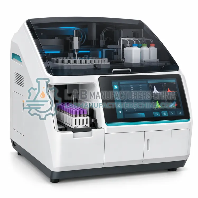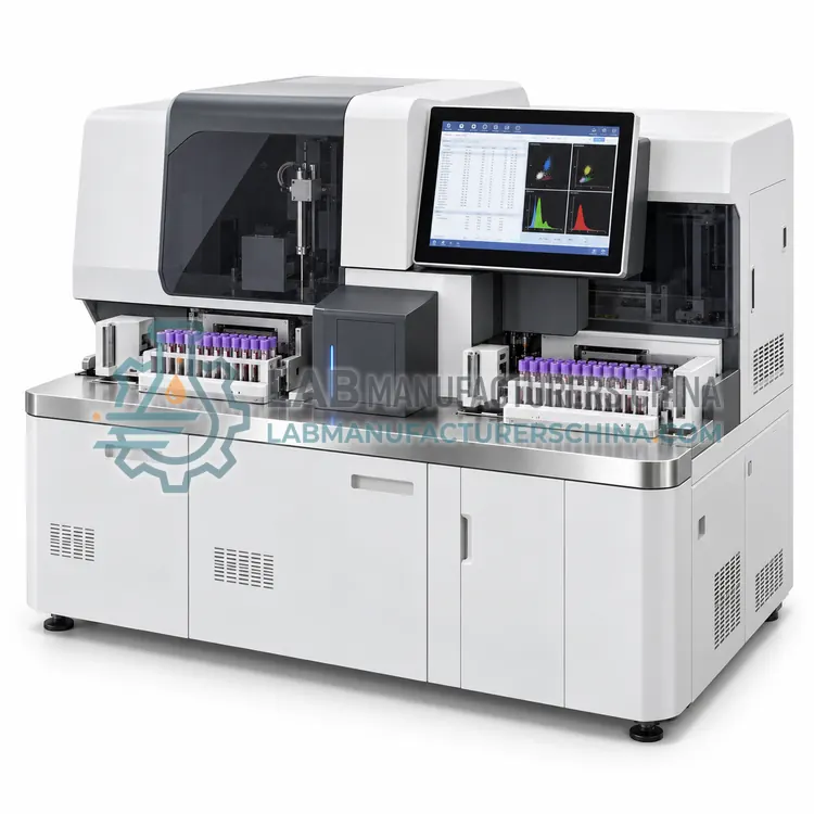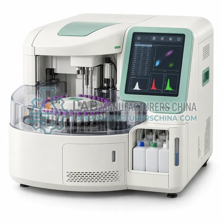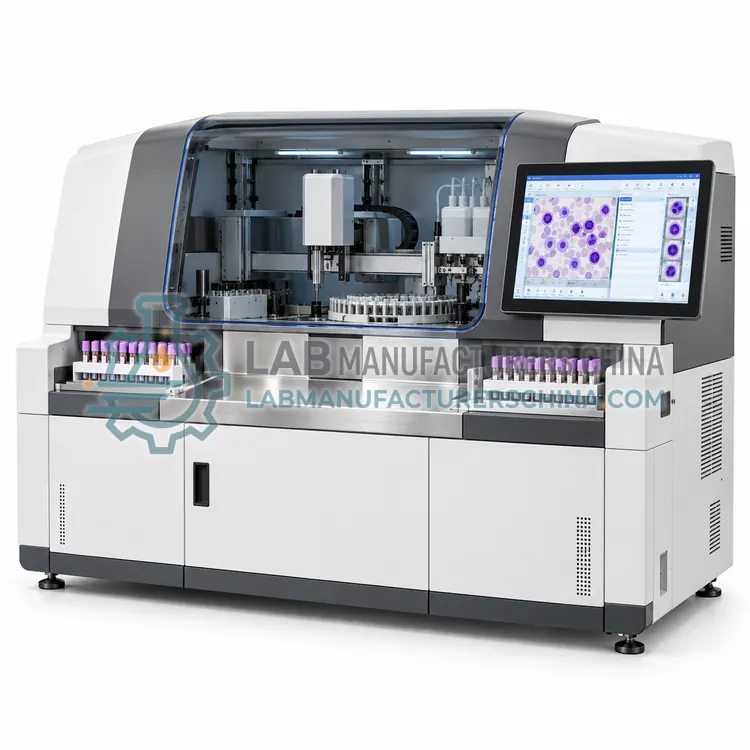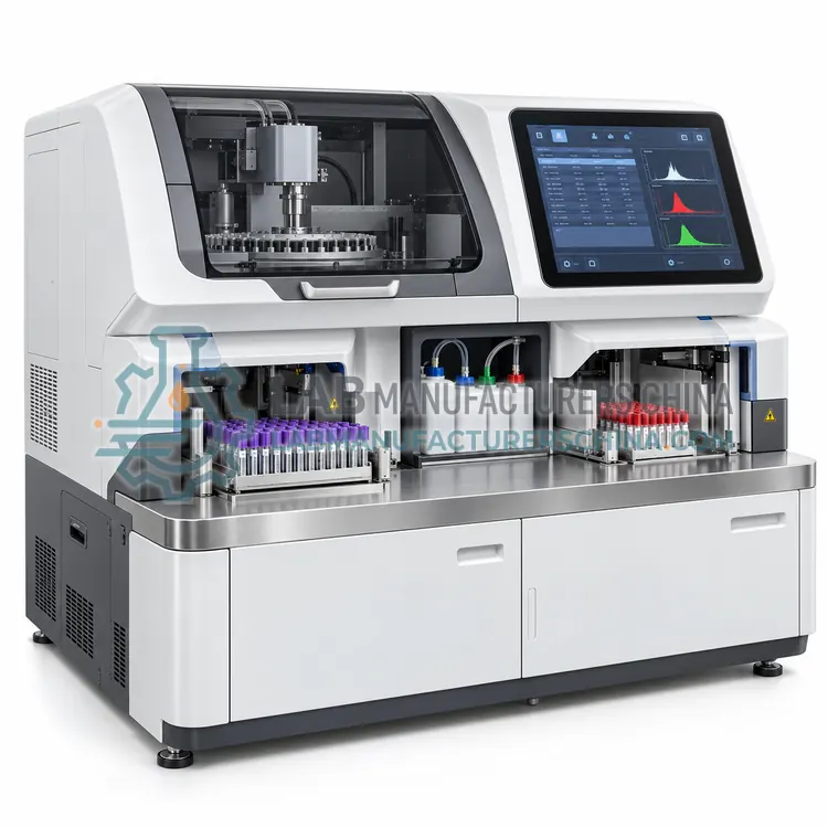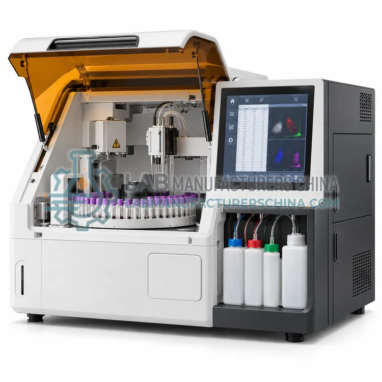
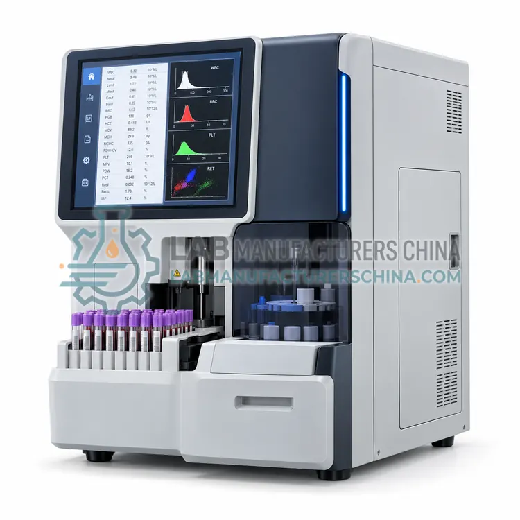
5-Part Hematology Analyzer with Reticulocyte Count
带网织红细胞计数的五分类血液分析仪
- SKU PAR-0003
- 5-Part Analyzers
- CE (to be confirmed)
- ISO 13485 manufacturing (to be confirmed)
- NMPA class II registration (to be confirmed)
Price on request — FOB / CIF quoted per order
Prices depend on configuration, quantity and destination port.
Key specifications
| Throughput | 60–80 samples/h with reticulocyte mode |
|---|---|
| Reticulocyte measuring range | 0.1–30 % |
| Reportable parameters | Reticulocyte %, absolute count, IRF, NRBC |
| Sample volume | 25 µL whole blood |
| Detection principle | Nucleic acid fluorescence plus impedance and scatter |
| Reticulocyte reagent stability | 60 days onboard at 20–25 °C protected from light |
Request a quote Download brochure · to be provided
By Lab Manufacturers China Technical TeamLast reviewed Standards: ISO 15189, CLSI H26-A2, CLSI H20-A2
What is the 5-Part Hematology Analyzer with Reticulocyte Count?
This analyzer adds a nucleic acid staining channel that quantifies reticulocytes and their maturity fractions alongside the five-part differential. It suits laboratories supporting hematology clinics, anemia workup and bone marrow transplant follow-up, where reticulocyte counts are requested daily.
What is the 5-Part Hematology Analyzer with Reticulocyte Count?
This analyzer adds a nucleic acid staining channel that quantifies reticulocytes and their maturity fractions alongside the five-part differential. It suits laboratories supporting hematology clinics, anemia workup and bone marrow transplant follow-up, where reticulocyte counts are requested daily.
Overview
Reticulocytes are young red cells that still contain residual RNA, so they can be distinguished from mature red cells by a dye that stains nucleic acids. In this analyzer a separate channel incubates the sample with the reticulocyte reagent, then measures fluorescence at high angle and forward scatter in the flow cell. The software reports reticulocyte percentage and absolute count, the immature reticulocyte fraction from the fluorescence intensity distribution, and derived indices such as corrected reticulocyte count. Because the fluorescent dye also labels white cells, an optional nucleated red cell count is usually included. Total throughput is slightly lower when the reticulocyte mode runs because of the extra incubation, typically 60 to 80 samples per hour, and the reagent must be protected from light and used within its stated stability.
How does it work?
These analyzers combine impedance counting with a flow cytometry channel. A diluted and stained sample passes a sheath flow cell where hydrodynamic focusing aligns cells in single file. A laser beam strikes each cell, and three signals are recorded: forward scatter, which reflects cell size; side scatter, which reflects internal granularity and complexity; and fluorescence from a dye that stains nucleic acids and cell membranes. Clustering algorithms in the instrument software separate the signals into five leukocyte populations, and the same data support flags for band forms, blasts, atypical lymphocytes and nucleated red cells. Impedance channels measure red cells, platelets and a second white cell count for comparison. Hemoglobin is measured photometrically after lysis. Because the flow channel provides much more information than a size histogram, the analyzer can also report reticulocytes, immature platelet fraction and body fluid cell counts when the corresponding reagent and software options are installed.
What are the key features?
- Dedicated fluorescence channel for reticulocyte counting
- Immature reticulocyte fraction derived from fluorescence intensity
- Nucleated red cell count from the same stained channel
- Five-part differential with blast and immature cell flags
- Automatic calculation of corrected and absolute reticulocyte counts
- Reagent stability monitoring with light-protected packaging
What are the technical specifications?
A typical 5-part hematology analyzer with reticulocyte count configuration offers throughput of 60–80 samples/h with reticulocyte mode, reticulocyte measuring range of 0.1–30 %, reportable parameters of Reticulocyte %, absolute count, IRF, NRBC, sample volume of 25 µL whole blood, detection principle of Nucleic acid fluorescence plus impedance and scatter and reticulocyte reagent stability of 60 days onboard at 20–25 °C protected from light. Values are indicative of the product class; the exact figures for each configuration are confirmed on the quotation and the calibration documents supplied with the instrument.
Which applications is it used for?
The 5-Part Hematology Analyzer with Reticulocyte Count is used in anemia classification and monitoring, bone marrow transplant engraftment follow-up, hemolytic disease assessment and hematology outpatient clinics. Across the wider 5-Part Analyzers class, laboratories also deploy it for hospital routine and emergency blood counts, neutropenia and chemotherapy monitoring and leukemia and lymphoma screening.
Which standards and certifications apply?
Relevant standards and methods include ISO 15189, CLSI H26-A2, CLSI H20-A2, CLSI EP05, CLSI H21, IEC 61010-1 and IEC 61010-2-101. Certification candidates for this model are CE, ISO 13485 manufacturing and NMPA class II registration; each is to be confirmed for the specific configuration before shipment, and certificates are supplied only where they have been issued.
Install the analyzer on a stable bench with a dedicated grounded outlet, controlled room temperature of about 15 to 30 degrees Celsius and low dust. Verify performance at installation with precision, carryover, linearity and method comparison studies, including a specific check of the differential against manual microscopy on abnormal samples. Run three levels of control each day, join an external quality assessment scheme, and review flag rates regularly, because an excessive flag rate increases manual reviews and delays reporting. Clean the flow cell, probes and waste lines according to the maintenance plan, protect the laser and optics from vibration, and keep sheath fluid and cleaning reagents within their expiry dates. Handle all blood samples as potentially infectious.
How do I select the right 5-Part Hematology Analyzer with Reticulocyte Count?
Order this variant when reticulocyte counts are requested several times daily, or when the laboratory serves a hematology service that relies on immature reticulocyte fraction for engraftment and treatment decisions. In the RFQ confirm the reticulocyte precision at low counts, whether nucleated red cell counting is included, and the onboard stability of the staining reagent. Check that the laboratory has a flow cytometric reference method available for validation.
How does it compare with other 5-Part Analyzers variants?
Specify a 5-part analyzer when clinicians act on the differential, for example to follow neutropenia during chemotherapy, to screen for leukemia, or to manage pediatric infections. Compare throughput, which ranges from about 60 to more than 120 samples per hour, and the first-result time for emergency work. Check which optional modes are included: reticulocyte count, nucleated red cell count, immature platelet fraction and body fluid analysis all require dedicated reagents and software licences. Confirm the minimum sample volume for pediatric samples and the linearity at low white cell counts. Ask about the flagging performance for blasts, the review rules the software applies, and whether a slide maker or digital morphology review is supported. Confirm LIS connectivity, reagent openness, calibration interval of the flow channel, and local service support.
Full specifications
| Throughput | 60–80 samples/h with reticulocyte mode |
|---|---|
| Reticulocyte measuring range | 0.1–30 % |
| Reportable parameters | Reticulocyte %, absolute count, IRF, NRBC |
| Sample volume | 25 µL whole blood |
| Detection principle | Nucleic acid fluorescence plus impedance and scatter |
| Reticulocyte reagent stability | 60 days onboard at 20–25 °C protected from light |
| Quality control | Three levels including a reticulocyte control |
| Data storage | 30,000 patient records |
| Interfaces | LAN with HL7 or ASTM, USB |
Indicative values for this product class. Final specifications are confirmed on the quotation and in the calibration documents supplied with the instrument.
Applications
- Anemia classification and monitoring
- Bone marrow transplant engraftment follow-up
- Hemolytic disease assessment
- Hematology outpatient clinics
Key features
- Dedicated fluorescence channel for reticulocyte counting
- Immature reticulocyte fraction derived from fluorescence intensity
- Nucleated red cell count from the same stained channel
- Five-part differential with blast and immature cell flags
- Automatic calculation of corrected and absolute reticulocyte counts
- Reagent stability monitoring with light-protected packaging
Frequently asked questions about 5-Part Hematology Analyzer with Reticulocyte Count
The immature reticulocyte fraction reflects the proportion of the youngest reticulocytes with the most residual RNA. It rises early after marrow recovery, for example following chemotherapy or transplantation, and is therefore used to monitor engraftment and marrow response before the total reticulocyte count changes appreciably.
No, most analyzers aspirate from the same EDTA tube used for the blood count and run the reticulocyte channel as an additional measurement on the same or a repeated aspiration. Some models need a slightly larger sample volume in that mode, which should be checked for pediatric collection tubes.
Validation compares the analyzer result with a reference method such as manual counting of supravitally stained films or a flow cytometric reference procedure, across low, normal and high values. Laboratories should document bias and precision and define review limits for discordant results.
A 5-part differential reports neutrophils, lymphocytes, monocytes, eosinophils and basophils separately rather than as grouped populations. This supports monitoring of neutropenia, allergic and parasitic conditions, and abnormal white cell patterns, and the underlying flow cytometry data also allow flags for immature cell populations.
No. Analyzers flag many abnormalities, but morphological assessment of blasts, dysplastic cells, malaria parasites and red cell shape still needs a stained film and microscopy. Most laboratories review films for a defined proportion of samples or whenever specific flags appear, as part of their validation and review rules.
Reticulocyte and nucleated red cell counts are useful for anemia assessment, the immature platelet fraction helps distinguish platelet destruction from reduced production, and body fluid modes support cerebrospinal and synovial fluid cell counts. Each module needs dedicated reagent and software, so order only what the clinical service will use.
Related models