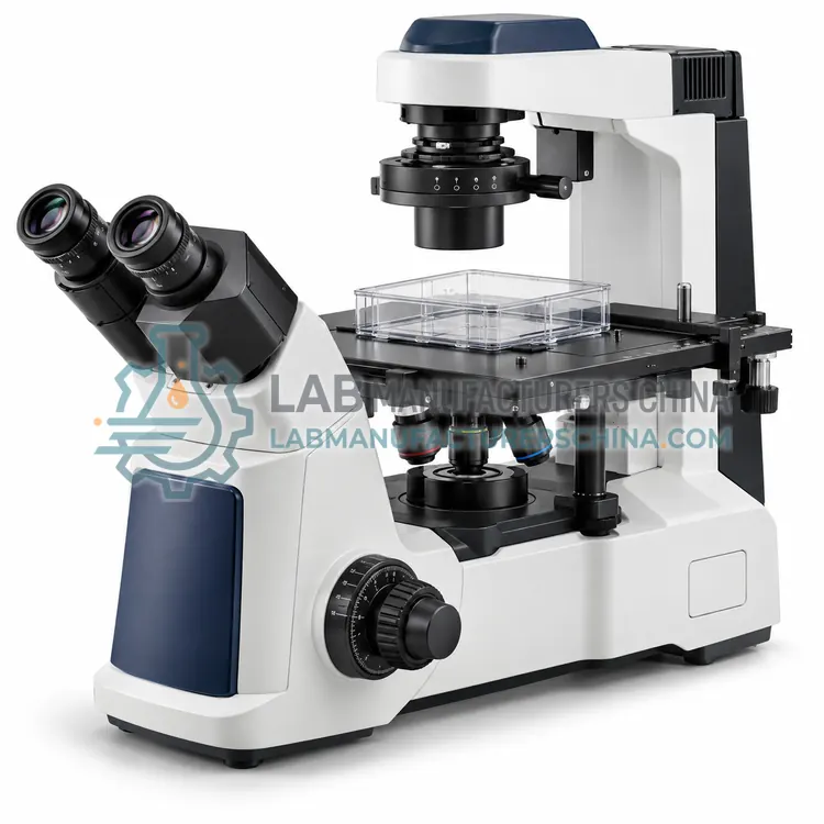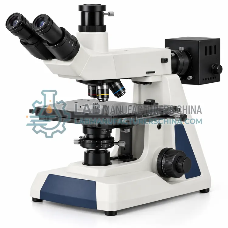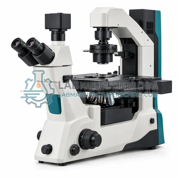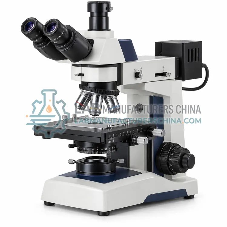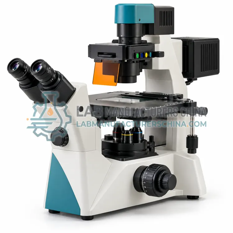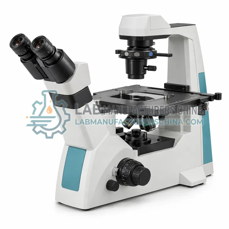
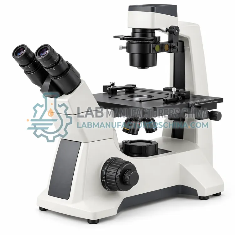
Routine Inverted Microscope
常规倒置显微镜
- SKU INM-0001
- Inverted Microscopes
- CE (to be confirmed)
- ISO 9001 manufacturing (to be confirmed)
Price on request — FOB / CIF quoted per order
Prices depend on configuration, quantity and destination port.
Key specifications
| Optical system | Infinite corrected |
|---|---|
| Magnification range | 40×–400× |
| Objectives | 4×, 10×, 20×, 40× long working distance |
| Eyepiece | 10× widefield |
| Working distance | Up to 6 mm |
| Stage | Mechanical, for flasks and dishes |
Request a quote Download brochure · to be provided
By Lab Manufacturers China Technical TeamLast reviewed Standards: ISO 8036, ISO 19012-1, ISO 9345-1
What is the Routine Inverted Microscope?
A routine inverted microscope provides basic brightfield and transmitted-light observation of specimens in flasks and dishes. It is an economical and reliable workhorse for daily cell-culture checks, routine screening and general laboratory examination.
What is the Routine Inverted Microscope?
A routine inverted microscope provides basic brightfield and transmitted-light observation of specimens in flasks and dishes. It is an economical and reliable workhorse for daily cell-culture checks, routine screening and general laboratory examination.
Overview
A routine inverted microscope uses a simple transmitted-light optical path with the objective below the stage. It typically offers brightfield and often a basic phase contrast option, fixed or limited magnification and a mechanical stage for positioning culture vessels. The stand is designed to hold flasks and dishes securely, and the working distance accommodates the vessel base. Illumination is by a long-life light-emitting diode with adjustable brightness. Controls are straightforward, so routine checks of cell confluency, contamination and morphology can be performed quickly. The design prioritises reliability and low cost over advanced imaging. A trinocular port is available on some models.
How does it work?
An inverted microscope reverses the usual arrangement so the objective looks upward from beneath the stage while the light source and condenser sit above. The specimen remains in a flask, dish or multi-well plate, so it need not be transferred to a slide. Long working distance objectives focus through the plastic or glass base of the vessel. Transmitted brightfield and phase contrast provide morphology, while episcopic illumination through the objective allows fluorescence. Ambient conditions can be controlled with heated stages and carbon dioxide chambers. The design supports observations of living material, micromanipulation and time-lapse imaging without disturbing the specimen.
What are the key features?
- Transmitted brightfield with optional phase contrast
- Objective below the stage for vessel-based samples
- Mechanical stage suited to flasks and dishes
- Long-life LED illumination with brightness control
- Simple controls for rapid routine checks
What are the technical specifications?
A typical routine inverted microscope configuration offers optical system of Infinite corrected, magnification range of 40×–400×, objectives of 4×, 10×, 20×, 40× long working distance, eyepiece of 10× widefield, working distance of Up to 6 mm and stage of Mechanical, for flasks and dishes. Values are indicative of the product class; the exact figures for each configuration are confirmed on the quotation and the calibration documents supplied with the instrument.
Which applications is it used for?
The Routine Inverted Microscope is used in routine cell culture checks, confluency and morphology assessment, contamination monitoring and general laboratory examination. Across the wider Inverted Microscopes class, laboratories also deploy it for live-cell and tissue culture microscopy, cell biology and stem-cell research and in vitro fertilisation and embryo work.
Which standards and certifications apply?
Relevant standards and methods include ISO 8036, ISO 19012-1, ISO 9345-1, ISO 8255-1 and IEC 61010-1. Certification candidates for this model are CE and ISO 9001 manufacturing; each is to be confirmed for the specific configuration before shipment, and certificates are supplied only where they have been issued.
Keep the vessel base clean and free of condensation, which scatters light and blurs the image. For phase contrast, centre the annulus with the matching objective. Maintain temperature and gas conditions for live cells, and minimise light exposure to reduce phototoxicity and bleaching. Long working distance objectives are exposed to the sample and medium, so clean them gently and avoid immersion of non-immersion optics. Calibrate measurements for each objective and vessel thickness, and record the setup with images. Service the stage and focus mechanisms regularly for stable time-lapse work.
How do I select the right Routine Inverted Microscope?
Choose a routine inverted microscope for daily culture monitoring where basic brightfield and optional phase contrast suffice. Confirm the stage accepts the flasks or dishes used and that the working distance is adequate. If fluorescence, micromanipulation or time-lapse imaging is planned, consider a more capable configuration.
How does it compare with other Inverted Microscopes variants?
Choose an inverted microscope whenever the specimen must stay in its vessel, especially living cells. Decide the observation method first: brightfield for stained or dense samples, phase contrast for unstained living cells, and fluorescence for labelled molecules. Confirm the working distance suits the vessel base and whether an environmental chamber or heated stage is required. For multi-well plates and automated imaging, a motorised stage reduces handling. For wafer or component inspection, an inverted metallurgical version with reflected illumination is used. Match objectives, condenser and camera to the required resolution.
Full specifications
| Optical system | Infinite corrected |
|---|---|
| Magnification range | 40×–400× |
| Objectives | 4×, 10×, 20×, 40× long working distance |
| Eyepiece | 10× widefield |
| Working distance | Up to 6 mm |
| Stage | Mechanical, for flasks and dishes |
| Illumination | 3 W LED, adjustable |
| Condenser | Long working distance, above stage |
| Focus adjustment | Coarse and fine |
| Power supply | 100–240 V AC |
Indicative values for this product class. Final specifications are confirmed on the quotation and in the calibration documents supplied with the instrument.
Applications
- Routine cell culture checks
- Confluency and morphology assessment
- Contamination monitoring
- General laboratory examination
Key features
- Transmitted brightfield with optional phase contrast
- Objective below the stage for vessel-based samples
- Mechanical stage suited to flasks and dishes
- Long-life LED illumination with brightness control
- Simple controls for rapid routine checks
Frequently asked questions about Routine Inverted Microscope
Many routine inverted microscopes offer phase contrast as an option with one or two phase objectives and a matching annulus. It is valuable for unstained cells, so check that the required phase objectives and condenser fit the model before purchase.
Most routine checks use 4× or 10× to survey a flask and 20× or 40× to inspect individual cells. Total magnifications from 40× to 400× cover confluency, morphology and contamination checks without needing high-numerical-aperture oil objectives.
Some models include a trinocular port or an integrated camera for basic documentation, while others rely on eyepiece viewing only. If images are needed for records, confirm the camera port and camera resolution before choosing a model.
With the objective below, the specimen stays in its culture vessel and the space above the stage is free for medium, tools and perfusion. This allows living cells to be observed without transferring them to a slide, and gives room for micromanipulation and environmental control.
Yes. Episcopic illumination passes through the objective from below, so fluorescence filter cubes work as in an upright microscope. This allows live cells in dishes to be imaged for labelled molecules while remaining in culture conditions.
The objective must focus through the plate base, typically about 1 mm thick, without touching it. Working distances of several millimetres are common for inverted objectives, and the best value depends on the vessel, the required numerical aperture and whether tools must also access the sample.
Related models