Microplate Fluorescence Reader
荧光微孔板读数仪
- SKU FLU-0002
- Fluorescence
- CE (to be confirmed)
- ISO 9001 manufacturing (to be confirmed)
- IVD-compatible configuration (to be confirmed)
Price on request — FOB / CIF quoted per order
Prices depend on configuration, quantity and destination port.
Key specifications
| Plate formats | 6 to 1536 wells |
|---|---|
| Excitation wavelength range | 230–900 nm |
| Emission wavelength range | 280–900 nm |
| Detection modes | FI, TRF, FP, luminescence |
| Detector | photomultiplier tube |
| Temperature control | ambient +4 to 45 °C |
Request a quote Download brochure · to be provided
By Lab Manufacturers China Technical TeamLast reviewed Standards: USP <853> Fluorescence Spectroscopy, ASTM E579 Standard Practice for Limit of Detection of Fluorescence, ICH Q2(R2) Validation of analytical procedures
What is the Microplate Fluorescence Reader?
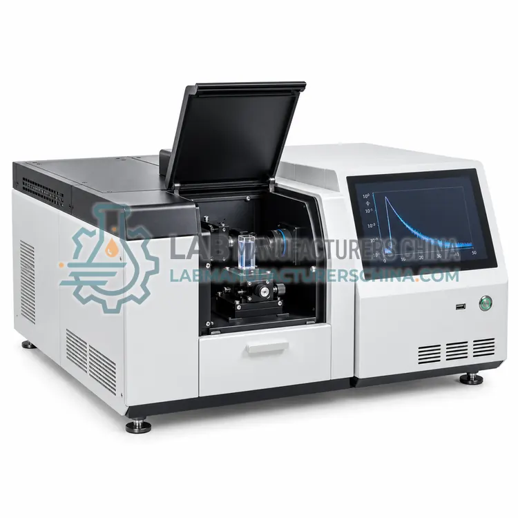
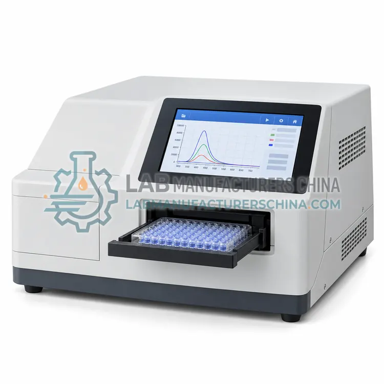
A microplate fluorescence reader measures fluorescence from samples in 96, 384 or 1536 well plates. It is the right choice for high-throughput screening, enzyme kinetics and immunoassays where many samples must be measured quickly and consistently.
What is the Microplate Fluorescence Reader?
A microplate fluorescence reader measures fluorescence from samples in 96, 384 or 1536 well plates. It is the right choice for high-throughput screening, enzyme kinetics and immunoassays where many samples must be measured quickly and consistently.
Overview
This variant replaces the single cuvette with a plate carrier and top or bottom optics that address each well in sequence. Excitation uses filters or monochromators, and emission is detected with a photomultiplier tube or CCD. Fluorescence intensity, time-resolved fluorescence and fluorescence polarization modes are common, and some models add luminescence and absorbance. Temperature control to 45 degrees Celsius, shaking and injectors support kinetic and cell-based assays. Cross-talk between wells is minimized by masking, and well-to-well repeatability is a key performance indicator. Robots and stackers automate plate handling for screening campaigns. Software provides plate layouts, standard curves, kinetic fits and export for downstream analysis, with audit trails available for regulated laboratories.
How does it work?
A fluorophore absorbs photons at an excitation wavelength, moves to an excited electronic state, loses some energy by non-radiative relaxation, and then emits photons at a longer wavelength when it returns to the ground state. The difference between excitation and emission maxima is the Stokes shift. A fluorescence spectrometer consists of an excitation source such as a xenon arc lamp, laser or LED, excitation and emission monochromators or filters, a sample holder for cuvettes, microplates or flow cells, and a detector such as a photomultiplier tube, avalanche photodiode or CCD. Because the detector is positioned at 90 degrees to the excitation beam, transmitted light is largely excluded and sensitivity can be orders of magnitude higher than absorption methods. Signal intensity depends on quantum yield, concentration, path length and quenching by oxygen, pH or matrix components.
What are the key features?
- Compatible with 6 to 1536 well microplates
- Fluorescence intensity, time-resolved and polarization modes
- Temperature control up to 45 degrees Celsius with shaking
- Filter or monochromator wavelength selection
- Automated plate stackers and robotic integration options
- Plate layout, standard curve and kinetic software with audit trail
What are the technical specifications?
A typical microplate fluorescence reader configuration offers plate formats of 6 to 1536 wells, excitation wavelength range of 230–900 nm, emission wavelength range of 280–900 nm, detection modes of FI, TRF, FP, luminescence, detector of photomultiplier tube and temperature control of ambient +4 to 45 °C. Values are indicative of the product class; the exact figures for each configuration are confirmed on the quotation and the calibration documents supplied with the instrument.
Which applications is it used for?
The Microplate Fluorescence Reader is used in high-throughput screening and hit confirmation, enzyme kinetics and inhibition assays, immunoassays and biomarker quantitation and cell viability and reporter gene assays. Across the wider Fluorescence class, laboratories also deploy it for pharmaceutical assay and impurity testing, nucleic acid and protein quantitation with fluorescent dyes and water quality monitoring of algae, chlorophyll and fluorescent tracers.
Which standards and certifications apply?
Relevant standards and methods include USP <853> Fluorescence Spectroscopy, ASTM E579 Standard Practice for Limit of Detection of Fluorescence, ICH Q2(R2) Validation of analytical procedures, ISO/IEC 17025 and IEC 61010-1. Certification candidates for this model are CE, ISO 9001 manufacturing and IVD-compatible configuration; each is to be confirmed for the specific configuration before shipment, and certificates are supplied only where they have been issued.
Install fluorescence instruments away from bright daylight and strong UV sources because stray light raises background. Xenon lamps produce ozone and heat and need adequate ventilation and lamp-hour tracking. Verify wavelength accuracy with reference standards and check sensitivity using the water Raman band or quinine sulfate in dilute sulfuric acid. Fluorescence is highly sensitive to dissolved oxygen, pH, temperature and quenching agents, so control and record these factors. Avoid photobleaching by limiting exposure of strongly fluorescent samples. Clean cuvettes and microplates must be free of fluorescent contaminants, and instrument qualification should follow IEC 61010-1 for safety and internal quality procedures for performance.
How do I select the right Microplate Fluorescence Reader?
Choose a microplate reader when sample count and speed drive the workflow, especially for screening, kinetic and cell-based assays. Specify plate formats, required detection modes, wavelength range, temperature and whether injectors or gas control are needed. Confirm well-to-well repeatability and cross-talk specifications, and check that the software supports your data export and compliance requirements.
How does it compare with other Fluorescence variants?
Select a fluorescence system by the sample format and the sensitivity required. Cuvette-based spectrofluorometers are best for method development and identity work because both excitation and emission wavelengths can be scanned. Microplate readers are the choice for high-throughput screening and enzyme assays in 96, 384 or 1536 wells. Time-resolved instruments measure fluorescence lifetimes and reject short-lived background for assays with long-lived labels. HPLC fluorescence detectors add selective quantitation to chromatographic separations. Portable filter fluorometers serve field water and tracer measurements where wavelength coverage matters less than speed. Compare excitation and emission ranges, stray-light rejection, detector type, minimum detectable concentration and software features such as spectral correction and blank subtraction.
Full specifications
| Plate formats | 6 to 1536 wells |
|---|---|
| Excitation wavelength range | 230–900 nm |
| Emission wavelength range | 280–900 nm |
| Detection modes | FI, TRF, FP, luminescence |
| Detector | photomultiplier tube |
| Temperature control | ambient +4 to 45 °C |
| Well-to-well repeatability | CV ≤ 2% |
| Read time for 96 wells | ≤ 15 s |
| Sensitivity | ≤ 1 fmol fluorescein per well |
| Shaking | adjustable orbital |
Indicative values for this product class. Final specifications are confirmed on the quotation and in the calibration documents supplied with the instrument.
Applications
- High-throughput screening and hit confirmation
- Enzyme kinetics and inhibition assays
- Immunoassays and biomarker quantitation
- Cell viability and reporter gene assays
Key features
- Compatible with 6 to 1536 well microplates
- Fluorescence intensity, time-resolved and polarization modes
- Temperature control up to 45 degrees Celsius with shaking
- Filter or monochromator wavelength selection
- Automated plate stackers and robotic integration options
- Plate layout, standard curve and kinetic software with audit trail
Frequently asked questions about Microplate Fluorescence Reader
Fluorescence polarization measures how much the polarization of emitted light is retained, which depends on how fast the labeled molecule tumbles in solution. It is used for binding assays, protease assays and molecular interaction studies because it reports on molecular size without separating bound and free label.
Cross-talk is limited by masking the optical path so only the target well is illuminated and read, by selecting plate materials with low fluorescence, and by using black plates for fluorescence assays. Well-to-well spacing, plate flatness and optics alignment all affect cross-talk, which is usually specified as a percentage of the signal.
Yes, most plate readers with temperature control hold 37 degrees Celsius within a fraction of a degree and support shaking and repeated reads for kinetics. For cell-based work, some models add carbon dioxide and humidity control. Confirm temperature stability, read interval and evaporation control for the plate type you use.
Fluorescence measures emitted photons against a dark background at 90 degrees to the excitation beam, so even a small number of emitted photons is detectable. Absorbance measures a small difference between two large light intensities. This signal-to-noise advantage lets fluorescence reach picomolar or lower concentrations that absorption methods cannot resolve.
Quenching reduces fluorescence intensity through collisions, energy transfer, dissolved oxygen or absorption of emitted light by the matrix. Control it by deoxygenating with nitrogen, matching solvent and pH to the validated method, diluting the matrix, and correcting with internal standards. Heavy atoms and colored compounds are common quenchers that must be identified during method development.
Standard cuvette instruments are designed for clear solutions, but front-face and solid-sample holders allow powders, films and turbid suspensions to be measured with reduced inner-filter effects. Integrating spheres improve reproducibility for scattering solids. For quantitative work, validated methods and reference materials for the specific matrix are essential because scattering complicates the relationship between intensity and concentration.
Related models